
|
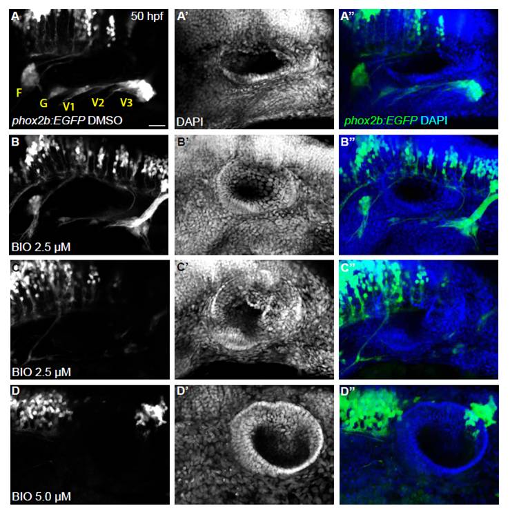
Fig. S9 Overactivation of Wnt signaling severely reduces or blocks EB ganglia formation. Confocal projection images of the otic vesicle and EB ganglia at 50 hpf. Tg(phox2b:EGFP)w37 embryos were treated with 2.5 and 5.0 μM BIO beginning 11 hpf, then washed out at 24 hpf. Embryos were allowed to develop until 50 hpf, at which time they were assessed for EB ganglion formation by EGFP expression and for otic vesicle formation by DAPI labeling. (A-C) Whereas DMSO-treated control embryos had normal development of the otic vesicle and EB ganglia, approximately half of 2.5 μM BIO-treated embryos had severely reduced EB ganglia and the remaining half had no EBs. (D) BIO (5.0 μM)-treated embryos resulted in a complete loss of EBs. Scale bar: 25 μm.