
Fig. 1
- ID
- ZDB-IMAGE-120724-2
- Genes
- Publication
- Akle et al., 2012 - F-Spondin/spon1b Expression Patterns in Developing and Adult Zebrafish
- All Figures
- Figures for Akle et al., 2012
|
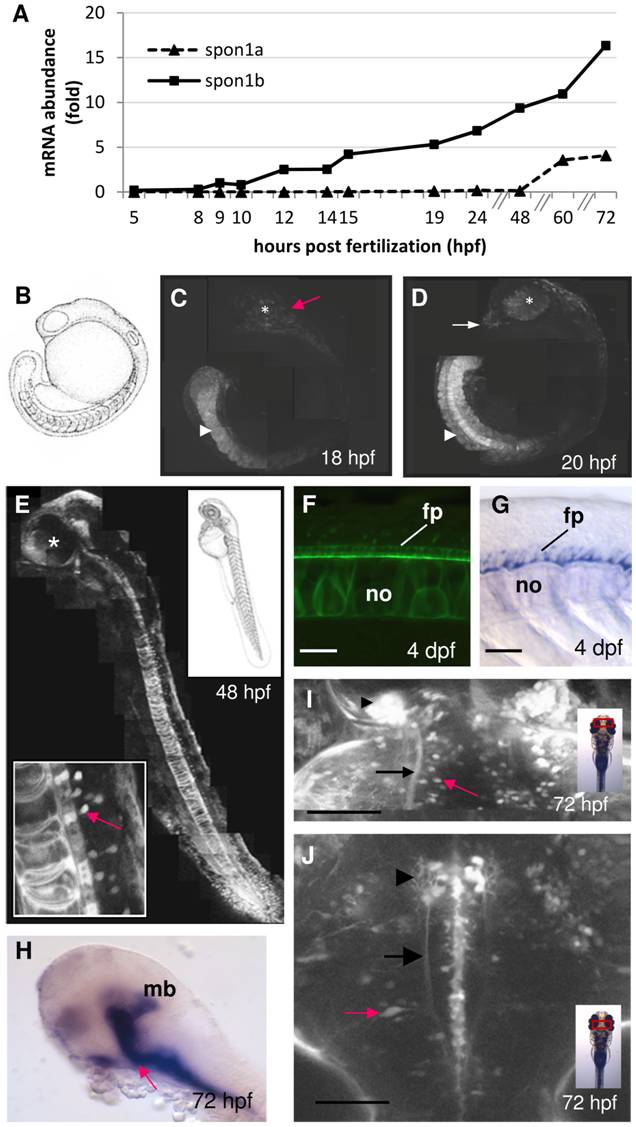
Fig. 1 F-spondin expression during early zebrafish development. A.
Onset of spon1a and spon1b expression during embryogenesis. Representative experiment showing mRNA abundance based on real-time RT-PCR (qPCR), with 1 fold corresponding to spon1b at 9 hpf. Zebrafish eggs fertilized at the same time (n>360) were sampled at intervals over a 72 h period (n = 30 embryos/larva per time point) and mRNA abundance for both genes was quantified in each same. B. Embryo schematic, 18 hpf. C-D. Spon1b:GFP expression in the tail bud, notochord and myotomes (arrowhead), brain areas (red arrow), olfactory bulbs (OB, white arrow) and retina (asterisk) at 18 hpf (B) and 20 hpf (D). Photomontages of confocal images. E. Spon1b:GFP expression pattern at 48 hpf. Photomontage of confocal images, sagittal view, asterisk: eye. Inset: developing motor neurons of the spinal cord (red arrow), dorsal to floor plate and notochord. F-G. The floorplate and notochord highlighted by spon1b:GFP (F, live image, 4 dpf) and in situ hybridization for spon1b mRNA (G, 4 dfp). Notochord (no), floorplate (fp). H. Spon1b expression in the flexural organ (arrow), and midbrain (mb). (in situ hybridization, 4 dpf). Rostral end to the left. I. Dorsal view of the telencephalon (Tel) and TeO border at 3 dpf, showing spon1b expression in habenula (arrowhead), in the fasciculus retroflexus (FR) emerging from it (black arrow), and in individual cells of the TeO (red arrow). Confocal z-stack image. J. Dorsal view of midbrain-hindbrain area at 3 dpf showing spon1b:GFP in the developing nMLF (arrowhead), in MLF projections (arrow) and in motor neurons of the reticular formation, including Mauthner cells (red arrow). Rostral end is up in I-J. Confocal z-stack image. Scale bars: F-G: 25 μm; I-J: 100 μm.