
Fig. 1
- ID
- ZDB-IMAGE-120724-18
- Genes
- Publication
- Flowers et al., 2012 - A zebrafish Notum homolog specifically blocks the Wnt/beta-catenin signaling pathway
- All Figures
- Figures for Flowers et al., 2012
|
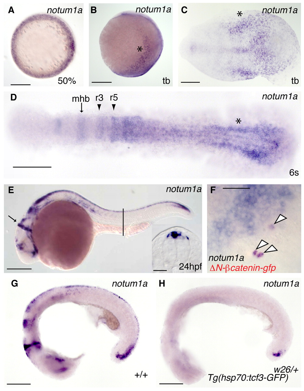
Fig. 1 Expression of zebrafish notum1a. (A-E) Expression of zebrafish notum1a. (A) At 50% epiboly, notum1a is expressed around the entire blastoderm margin. (B) A lateral view of notum1a expression at tail bud reveals expression in the lateral neural plate (asterisk). (C) A flat-mount of the tail bud displays notum1a expression in the posterior neural plate, particularly at the lateral edges (asterisk) and midline. (D) At six somites, as neurulation proceeds, lateral neural plate notum1a expression becomes more medially located. More anteriorly, notum1a begins to appear in the midbrain-hindbrain boundary (mhb, arrow) and rhombomeres 3 and 5 (r3, r5, arrowheads). (E) At 24 hpf, notum1a is expressed in the mhb (arrow) and in the dorsal neural tube, as revealed in a section through the trunk at the level of the yolk extension (line, inset). Scale bar: 50 μm. (F,G) Wnt/β-catenin signaling is needed for notum1a expression. (F) Mosaically expressed activated β-catenin (red) colocalizes with ectopic notum1a expression (arrowheads) at 80% epiboly. Scale bar: 50 μm. (G,H) Although normally expressed in heat-shocked non-transgenic embryos at 22 hpf (G), notum1a is absent in heat-shocked Tg(hsp70:tcf3-gfp)w26/+ embryos (H) except in small domain within the tail bud 2 hours post-heat shock. Scale bars in A-D,G,H: 200 μm.