
Image
|
Figure Caption
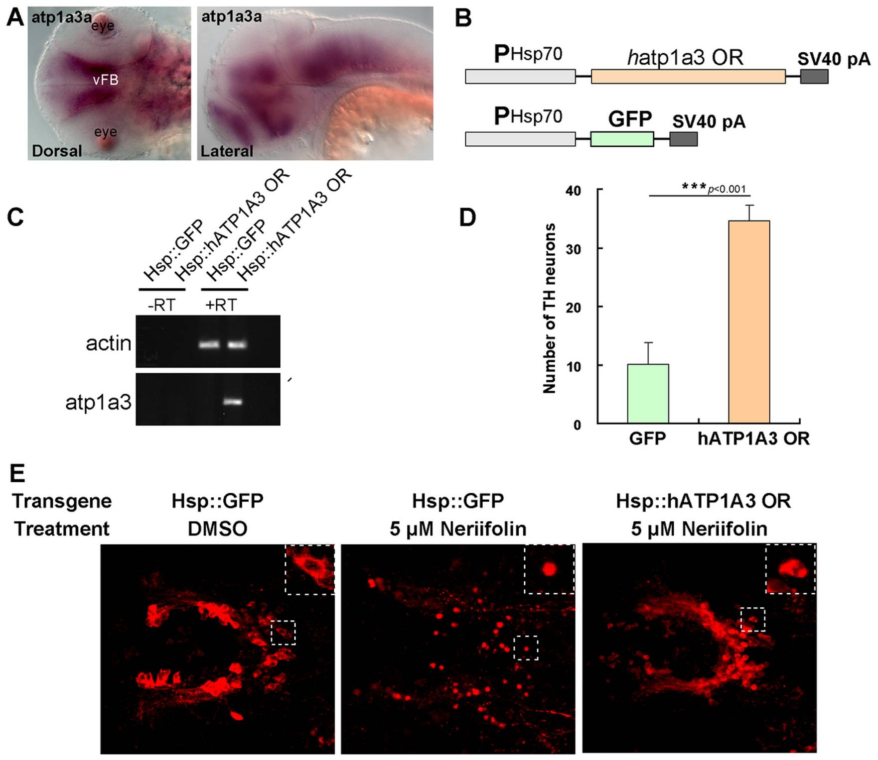
Fig. 2 Human atp1a3 rescues DA neurons in Neriifolin-treated embryos.
(A) The expression pattern of atp1a3a in wild-type embryos at 48 hpf. (B) The schematic diagram of the plasmid constructs used for the rescue experiments in zebrafish embryos. (C) RT-PCR detection of the expression of human atp1a3 in zebrafish embryos after injection and heat shock. (D–E) Quantification (D) and representative images (E) of VFB DA neurons in 5 μM Neriifolin-treated embryos that express either GFP or human atp1a3. Data are the averages ± SEM from 9 embryos in a single experiment that was repeated twice with similar results.
Acknowledgments
This image is the copyrighted work of the attributed author or publisher, and
ZFIN has permission only to display this image to its users.
Additional permissions should be obtained from the applicable author or publisher of the image.
Full text @ PLoS One