
Fig. 2
- ID
- ZDB-IMAGE-120427-1
- Genes
- Publication
- Chen et al., 2012 - Zebrafish agr2 is required for terminal differentiation of intestinal goblet cells
- All Figures
- Figures for Chen et al., 2012
|
Fig. 2
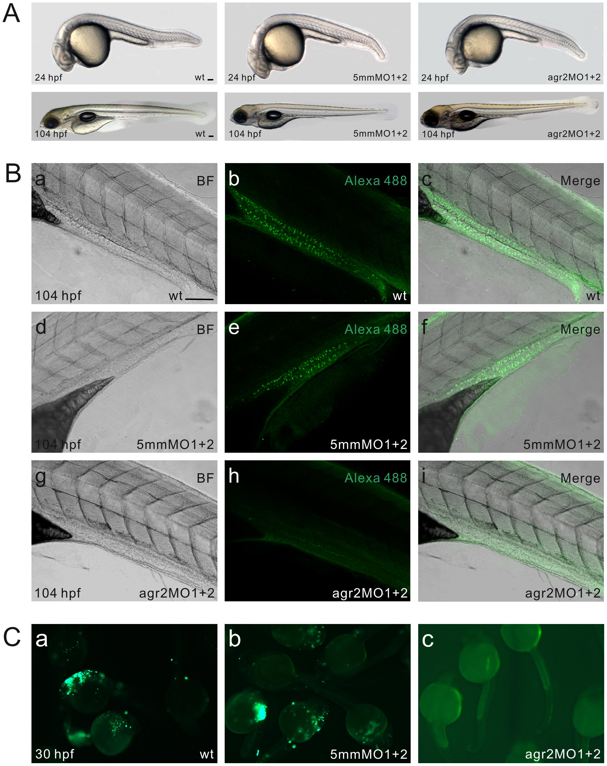
agr2 morpholino antisense oligomer knockdown analyses.
(A) Phenotype comparison among wild type, agr2–5 mmMO1 and 5 mmMO2-coinjected, and agr2-MO1 and agr2-MO2-coinjected embryos at 24 and 104 hpf. (B) Whole-mount immunohistochemistry demonstrates that coinjection of agr2-MO1 and agr2-MO2 prevents the synthesis of Agr2 protein in intestinal goblet cells. Confocal images of either wild type, agr2–5 mmMO1 and 5 mmMO2-coinjected, or agr2-MO1 and agr2-MO2-coinjected 104 hpf embryos were recorded under transmitted mode (a, d, g) or using 494/517 nm excitation/emission wavelengths (b, e, h). Merged images are shown (c, f, i). (C) Green fluorescence was not detected in agr2-MO1, agr2-MO2 and CMV-agr2-mo-GFP coinjected (c) 30 hpf embryos, whereas bright green fluorescence was observed in CMV-agr2-mo-GFP-injected (a) and agr2–5 mmMO1, agr2–5 mmMO2 and CMV-agr2-mo-GFP coinjected (b) 30 hpf embryos. Scale bars represent 100 μm.