
Fig. 2
- ID
- ZDB-IMAGE-120425-15
- Genes
- Publication
- Sultana et al., 2008 - Zebrafish early cardiac connexin, Cx36.7/Ecx, regulates myofibril orientation and heart morphogenesis by establishing Nkx2.5 expression
- All Figures
- Figures for Sultana et al., 2008
|
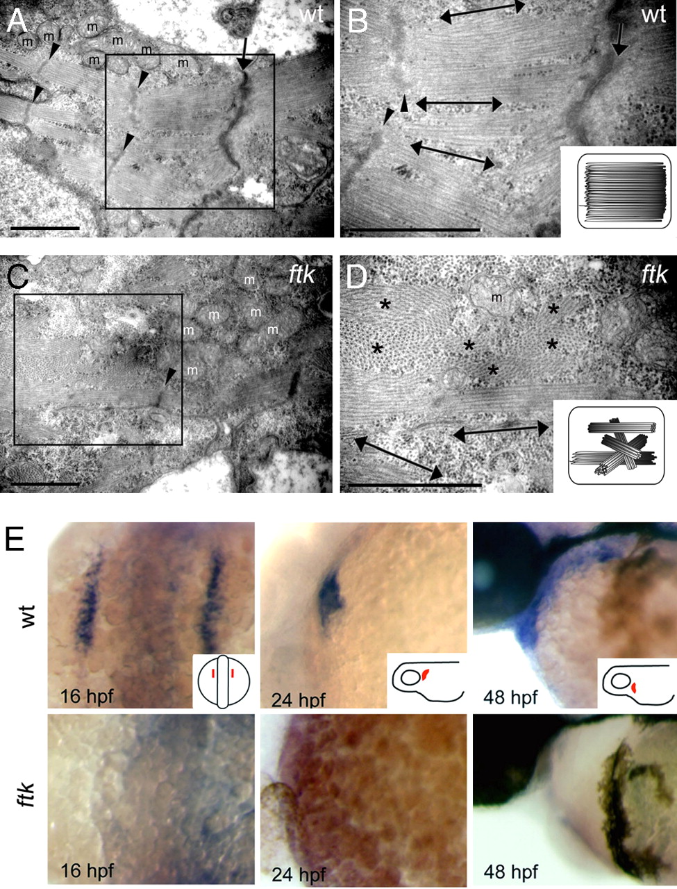
Fig. 2
ftk mutation primarily affected the myofibril organization and nkx2.5 expression. (A–D) Myofibril organization examined by TEM analysis. In wild-type embryos at 58 hpf (A and B), myofibrils assemble as thick parallel bundles with orientation toward the longitudinal direction of the cardiomyocyte (double-headed arrows). In contrast, myofibrils in the ftk heart (C and D) are oriented in irregular directions. Double-headed arrows and asterisks indicate the myofibril bundles sectioned parallel and perpendicular to the long axis of the cells, respectively. Arrowheads, Z-lines; arrows, cell–cell contact sites; m, mitochondria. (Scale bars: 500 nm.) (B and D Insets) Schematic drawings of myofibril organization. (E) Whole-mount in situ hybridization analysis of nkx2.5 expression. In wild-type zebrafish embryos (Upper) The expression is detected in the developing heart; however, it is not detectable in ftk mutants (Lower). Mutants at early stages were genotyped by RFLP analysis. (Insets) Orientations of embryos.