
Fig. 1
- ID
- ZDB-IMAGE-120425-14
- Publication
- Sultana et al., 2008 - Zebrafish early cardiac connexin, Cx36.7/Ecx, regulates myofibril orientation and heart morphogenesis by establishing Nkx2.5 expression
- All Figures
- Figures for Sultana et al., 2008
|
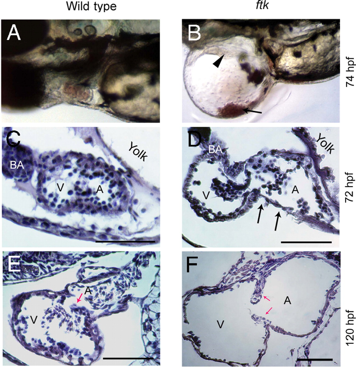
Fig. 1
Progressive heart malformation in zebrafish ftk mutant. (A and B) Gross heart morphology of ftk mutant. Wild-type (A) and ftk mutant embryos (B) at 74 hpf. Arrowhead indicates heart perforations in the ftk mutant; arrow points to blood cells accumulated in the pericardiac cavity. (C–F) Sagittal sections of the heart. At 72 hpf (C and D), a dilated atrium (A) and ventricle (V) are evident in the ftk mutant. The cardiac muscles appear to be thinner (arrows in D), and the dilation of heart chambers is under way. At 120 hpf (E and F), the heart chamber dilation becomes more severe in ftk mutants with thinner ventricular and atrial walls. Red arrows indicate the valve leaflets. (Scale bars: 150 μm.)