Fig. 3
Fig. 3
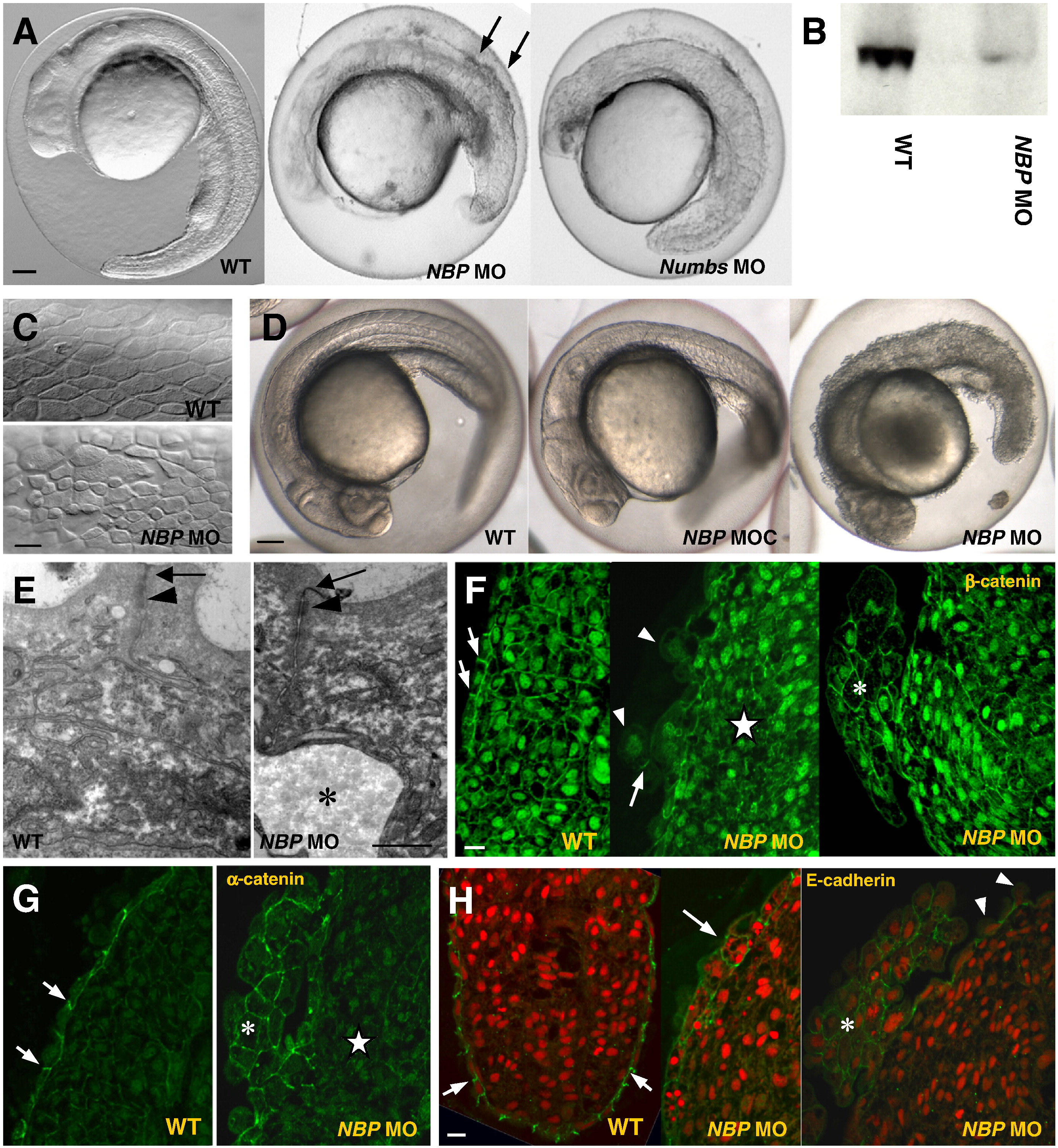
NBP and Numbs morphants show defects in development. Panels compare NBP morphants (NBP MO), NBP control morphants (NBP MOC) and Numbs morphants (Numbs MO) with wild-type embryos (WT). (A) 24 hpf embryos injected with either NBP MO or Numbs MO are delayed in growth and abnormally developed in comparison with wild-type embryos.Arrows point to dissociated cells in the dorsal region of the NBP morphants (lateral view, anterior towards the left). (B) Western blot analysis of protein extracts from 24hpf wild-type and NBP MO-injected embryos with the N1 polyclonal antibody against NBP. The wild-type extract gives a NBP-positive band of the expected size (99.57 kDa), whereas only a negligible amount of NBP is seen in extracts from the morphants. (C, E–H) Anomalous development of the epidermis. The epidermal cells of the NBP MO-injected embryos varied in size and shape, were separated by large intercellular spaces (C) and frequently were released from the surface (E, asterisk denotes the gap below the epidermal cells; arrowheads in F and H). Tumor-like structures emerged from the dorsal part of the embryo (asterisks in F, G, and H). They were composed of enlarged cells with round, β-catenin strongly positive nuclei (F) and with α-catenin (G), β-catenin (F) and E-cadherin (H) enriched in the cell–cell contacts. Stars point to inner tissue located just underneath the tumor-like structures where cells lack β-catenin in both nuclei and membranes (F) and α-catenin in membranes (G). Arrows point to junctions between neighboringepidermal cells enriched in β-catenin (F), α-catenin (G) and E-cadherin (H) in WT and to mislocalized E-cadherin in epidermal cells of NBP MO (H). Nuclei in H are stained by acridine orange. Scale bar: A, D = 100 μm, C, F–H = 20 μm, and E = 1 μm.
Reprinted from Developmental Biology, 365(1), Boggetti, B., Jasik, J., Takamiya, M., Strähle, U., Reugels, A.M., and Campos-Ortega, J.A., NBP, a zebrafish homolog of human Kank3, is a novel Numb interactor essential for epidermal integrity and neurulation, 164-174, Copyright (2012) with permission from Elsevier. Full text @ Dev. Biol.

