
|
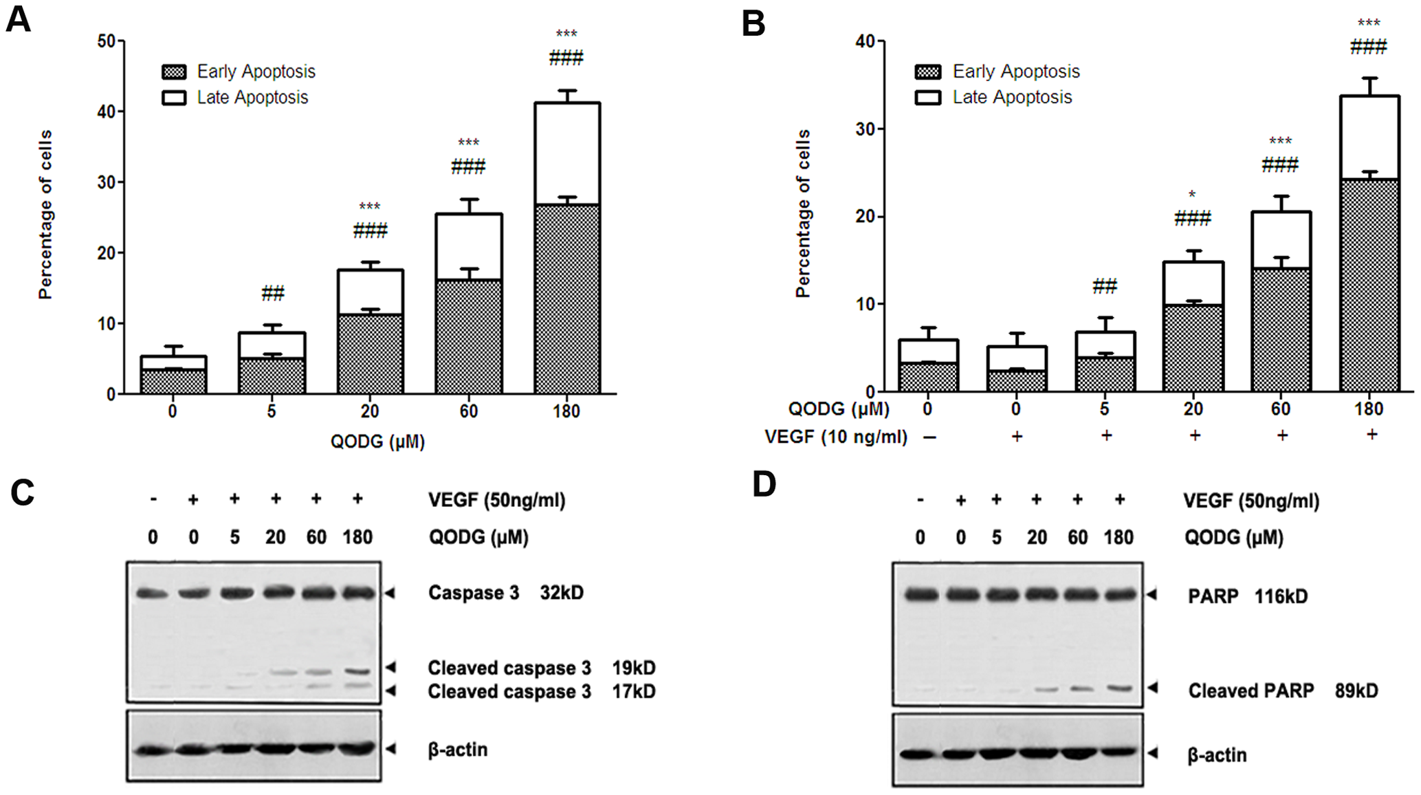
Fig. 11
QODG potentiates apoptosis in HUVECs in a dose-dependent manner.
(A–B) Relative percentages of early apoptotic cells (annexin-V+/PI-) and necrotic or late apoptotic cells (annexin-V+/PI+) were analyzed with one-way ANOVA followed by Tukey′s multiple comparison test. Cells receiving only DMSO (0.1%) served as a vehicle control. Data are expressed as percentages of the vehicle control (100%) in mean ± SD from three independent experiments. (A) The percentages of early apoptotic cells and necrotic or late apoptotic cells increased in a dose-dependent manner when HUVECs were treated without VEGF. #, the percentage of early apoptotic cells (annexin-V+/PI-) in QODG-treated group compared with that in the vehicle control group; ##, P<0.01 vs. vehicle control; ###, P<0.001 vs. vehicle control. *, the percentage of late apoptotic cells (annexin-V+/PI+) in QODG-treated group compared with that in the vehicle control group; ***, P<0.001 vs. vehicle control. (B) The percentages of early apoptotic cells and necrotic or late apoptotic cells increased in a dose-dependent manner when HUVECs were treated with VEGF. #, the percentage of early apoptotic cells (annexin-V+/PI-) in QODG-treated group compared with that in the VEGF-treated control group; ##, P<0.01 vs. VEGF-treated control; ###, P<0.001 vs. VEGF-treated control. *, the percentage of late apoptotic cells (annexin-V+/PI+) in QODG-treated group compared with that in the VEGF-treated control group; *, P<0.05 vs. VEGF-treated control; ***, P<0.001 vs. VEGF-treated control. (C–D) QODG induced caspase-3 activation and the cleavage of PARP from its intact form to its cleaved form. Proteins from HUVECs treated with or without VEGF (10 ng/mL) and DMSO (0.1%) or various concentrations of QODG (5, 20, 60, 180 μM) were analyzed by Western blotting analysis for cleaved caspase-3 and cleaved PARP.