
|
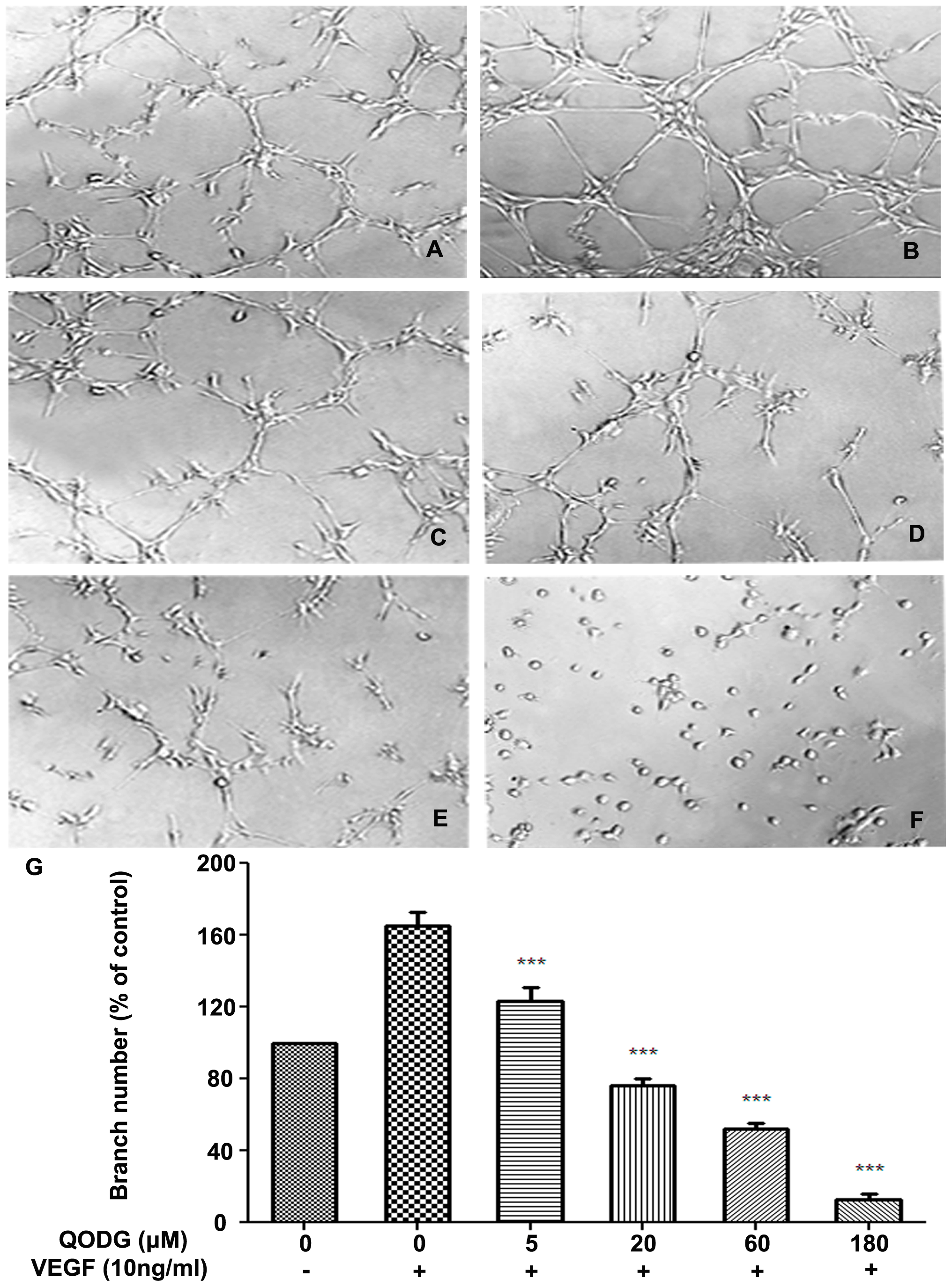
Fig. 7
QODG inhibits VEGF-induced capillary structure formation of endothelial cells on Matrigel.
QODG inhibited VEGF-induced tube formation of HUVECs. HUVECs were starved with ECGM containing 0.5% FBS, and then treated with DMSO (0.1%) or various concentrations of QODG (5, 20, 60, 180 μM). After that, cells were collected and placed in 24-well plates coated with Matrigel (4×10⁴ cells/well), followed by the activation of VEGF (10 ng/mL). After 6 h of incubation, images of the network-like structures of endothelial cells were taken using an inverted microscope (Olympus, Center Valley, PA, USA) (at 100×magnification), and branching points in different groups were quantified by manual counting. (A) HUVECs cultured on Matrigel were treated with only DMSO (0.1%). (B) HUVECs cultured on Matrigel were treated with VEGF (10 ng/mL) and DMSO (0.1%). (C–F) HUVECs cultured on Matrigel were treated with VEGF (10 ng/mL) and various concentrations of QODG (5, 20, 60, 180 μM). (G) Quantitative comparison of the numbers of branching points in different groups. Cells receiving only DMSO (0.1%) served as a vehicle control. Data are expressed as percentages of the vehicle control (100%) in mean ± SD from three independent experiments. ***, P<0.001 vs. VEGF-treated control. Scale bars, 100 μm.