
|
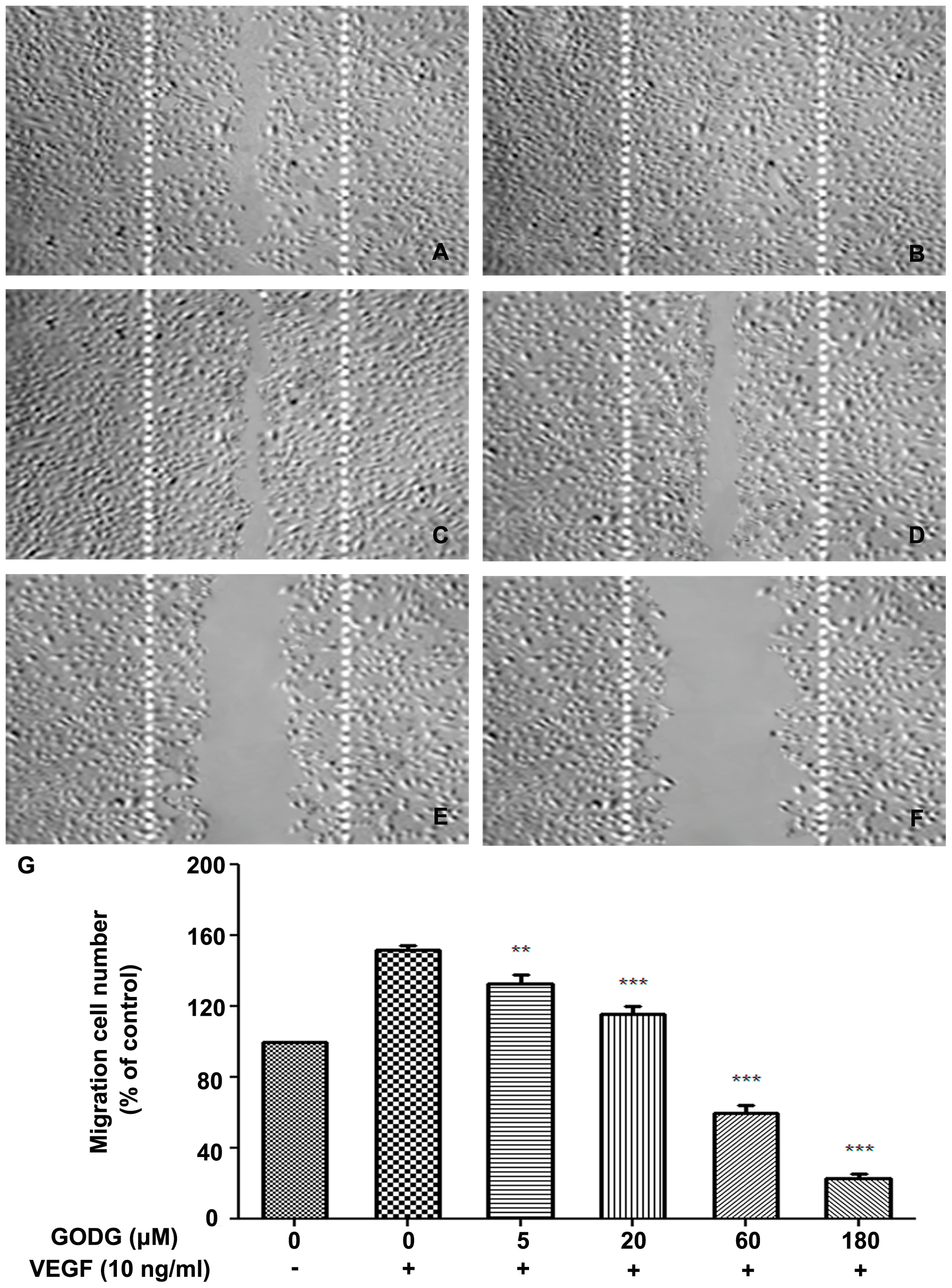
Fig. 6
QODG inhibits VEGF–induced chemotactic motility of endothelial cells.
QODG inhibited the migration of HUVECs. HUVECs were allowed to grow to full confluence in 6-well plates pre-coated with 0.1% gelatin and then starved with ECGM containing 0.5% FBS to inactivate cell proliferation. After that, cells were wounded with pipette and washed with PBS, then treated with or without VEGF (10 ng/mL) and DMSO (0.1%) or different concentrations of QODG (5, 20, 60, 180 μM) in ECGM containing 0.5% FBS. Images were taken using an inverted microscope (Olympus, Center Valley, PA, USA) (at 100×magnification) after 8 h of incubation, and migrated cells were quantified by manual counting. (A) Migration assay of HUVECs treated with only DMSO (0.1%). (B) Migration assay of HUVECs treated with VEGF (10 ng/mL) and DMSO (0.1%). (C–F) Migration assay of HUVECs treated with VEGF (10 ng/mL) and various concentrations of QODG (5, 20, 60, 180 μM). (G) Quantitative comparison of the numbers of migrated cells in different groups. Cells receiving only DMSO (0.1%) served as a vehicle control. Data are expressed as percentages of the vehicle control (100%) in mean ± SD from three independent experiments. **, P<0.01 vs. VEGF-treated control; ***, P<0.001 vs. VEGF-treated control. Scale bars, 100 μm.