
Fig. 5
- ID
- ZDB-IMAGE-120216-88
- Genes
- Publication
- Wu et al., 2011 - Ryanodine receptors, a family of intracellular calcium ion channels, are expressed throughout early vertebrate development
- All Figures
- Figures for Wu et al., 2011
|
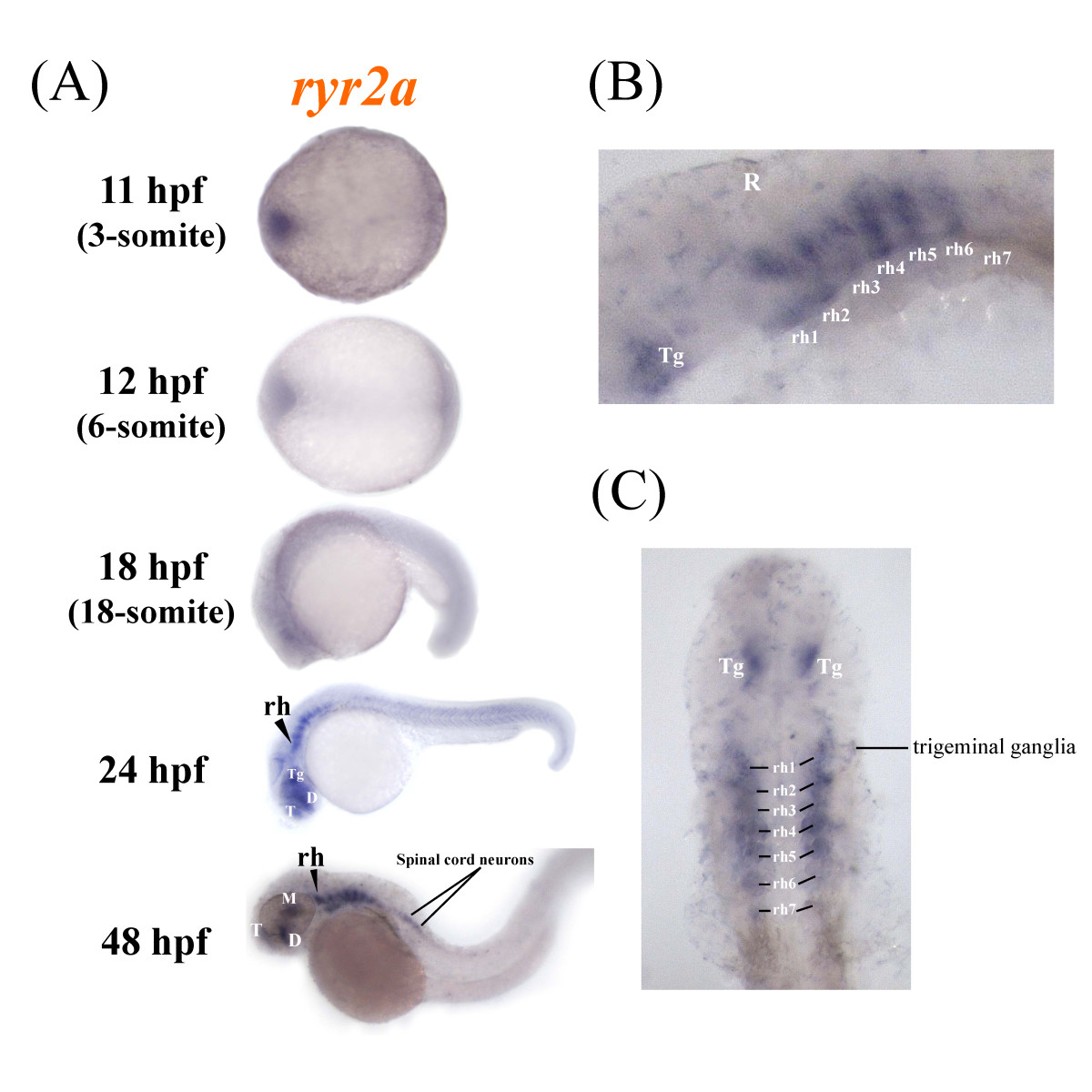
Fig. 5
Zebrafish ryr2a is expressed in the central nervous system. The spatial distribution of ryr2a mRNA in the zebrafish embryo was examined using whole mount in situ hybridisation. (a) Specific staining was observed initially in localised regions of the brain, specifically the telencephalon (T), diencephalon (D), mesencephalon (M), tegmentum (Tg) and rhomobomeres (rh) of whole mount embryos at 24 and 48 hpf. (b) Lateral and (c) dorsal views (eyes removed) of the brain at 48 hpf revealed that ryr2a expression was present in each of the seven rhombomere segments, the tegmentum, trigeminal ganglia and anterior portion of the spinal cord. Embryos are orientated so that anterior is to the left.