
Fig. S2
- ID
- ZDB-IMAGE-120216-106
- Publication
- Van Otterloo et al., 2012 - Novel Tfap2-mediated control of soxE expression facilitated the evolutionary emergence of the neural crest
- All Figures
- Figures for Van Otterloo et al., 2012
|
Fig. S2
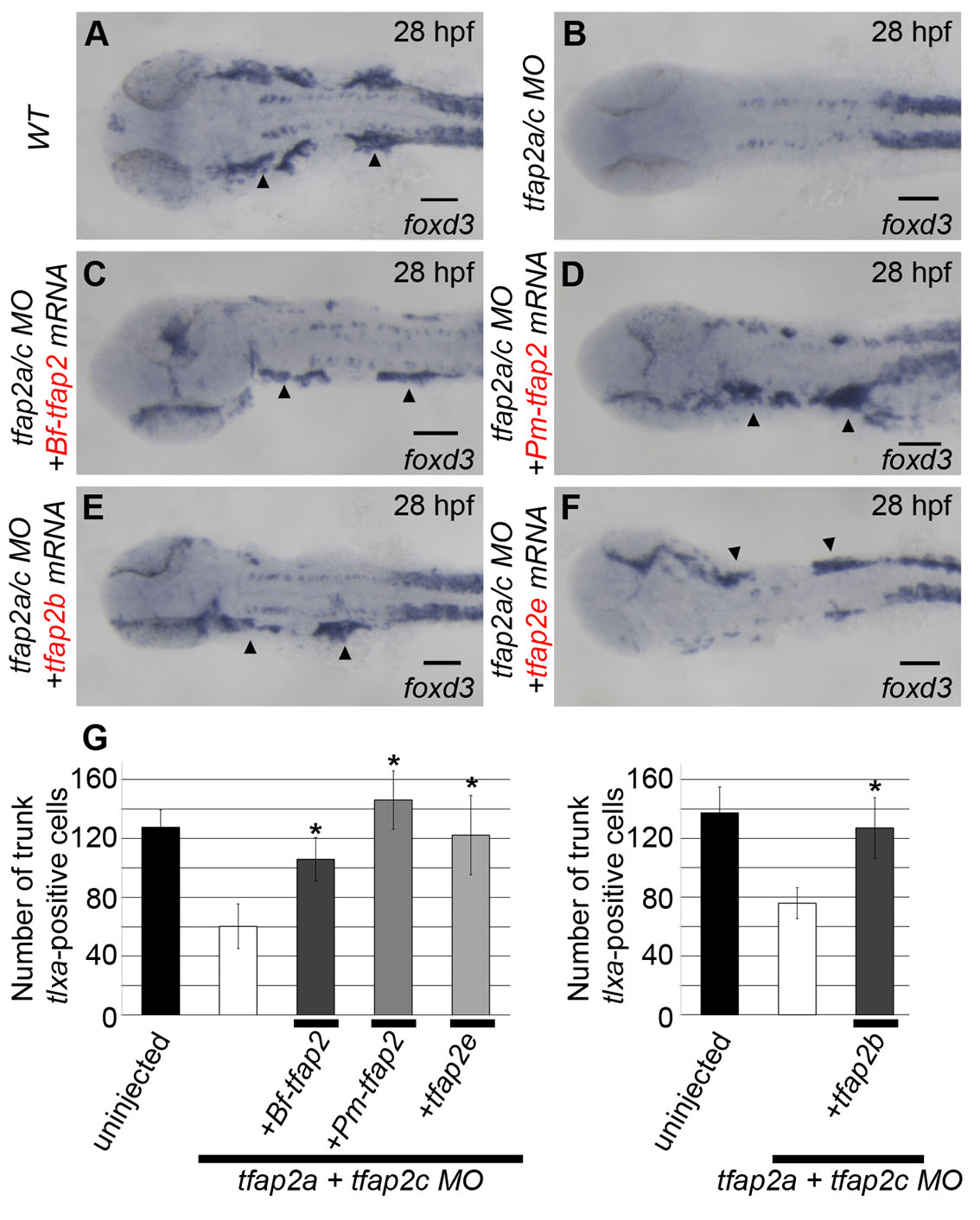
Additional neural crest derivatives rescued by tfap2 homologs. (A-F) All embryos are viewed dorsally and oriented with anterior to the left. (A) Uninjected embryos show clear foxd3 expression at 28 hpf marking the cranial ganglia (arrowheads, 23/23). (B) This expression is absent in embryos injected with MOs targeting tfap2a and tfap2c (0/24). Embryos injected with these MOs and subsequently injected with mRNA encoding (C) amphioxus tfap2 (29/35), (D) lamprey tfap2 (26/26), (E) zebrafish tfap2b (31/33) and (F) tfap2e (22/22) show restored foxd3 expression within the cranial ganglia on either one or both sides (mRNA was injected mosaically). (G) Graph showing cell counts of tlxa-positive cells at 22 hpf, a marker of Rohon-Beard and dorsal root ganglia sensory neuron precursor cells, found in the trunk of embryos injected similarly to those shown in A-F (n=10/group, *P<0.05 relative to tfap2a/tfap2c MO alone group). Of note, the incomplete absence of tlxa-positive cells in tfap2a/tfap2c MO-injected embryos is a previously described Tfap2-independent population (Li and Cornell, 2007).