
IMAGE
Fig. 2
- ID
- ZDB-IMAGE-120208-1
- Genes
- Antibodies
- Publication
- Du et al., 2012 - Differential regulation of epiboly initiation and progression by zebrafish Eomesodermin A
- All Figures
- Figures for Du et al., 2012
Image
|
Figure Caption
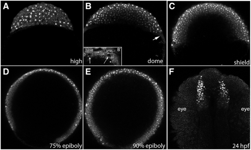
Fig. 2 Eomesa protein distribution. Confocal projections of embryos stained with anti-Eomesa antibody. (A-E) lateral views (F) dorsal view. Stages indicated in lower right. Arrow in (B) indicates the YSL. (B′) Inset shows YSL of embryo at sphere stage. Arrows indicate unstained YSL-nuclei that are surrounding by Eomesa positive YSL cytoplasm.
Figure Data
Acknowledgments
This image is the copyrighted work of the attributed author or publisher, and
ZFIN has permission only to display this image to its users.
Additional permissions should be obtained from the applicable author or publisher of the image.
Reprinted from Developmental Biology, 362(1), Du, S., Draper, B.W., Mione, M., Moens, C.B., and Bruce, A.E., Differential regulation of epiboly initiation and progression by zebrafish Eomesodermin A, 11-23, Copyright (2012) with permission from Elsevier. Full text @ Dev. Biol.