
|
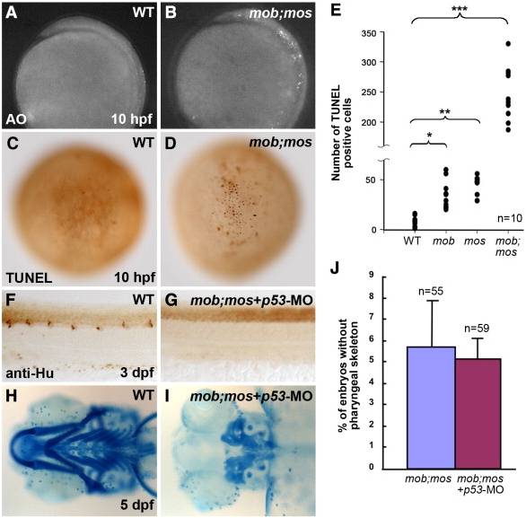
Fig. 2 Apoptosis is not the primary cause of the mob;mos double mutant phenotype. (A, B) Lateral view of acridine orange (AO) staining of wild-type (WT) (A) and mob;mos (B) embryos at 10 hpf. (C, D) Dorsal view of TUNEL assay in WT (C) and mob;mos (D) embryos revealing more apoptotic cells in the neural plate region of mob;mos embryos at 10 hpf. (E) Quantification of TUNEL positive cells in the neural plate region of WT, mob, foxd3-MO-injected, and foxd3-knockdown mob embryos. Each spot represents one embryo. n signifies the number of embryos counted. Error bars represent standard deviation. Data analyzed by Student′s t-test, *P < 0.05, **P < 0.01, and ***P < 0.001. (F–J) Suppression of apoptosis by knock-down of p53 does not rescue the phenotype in mob;mos embryos. Lateral view of anti-Hu immunostaining at 3 dpf in WT (F) and mob;mos (G) embryos injected with p53 MO. Ventral view of Alcian blue staining of the head at 5 dpf of WT (H) and mob;mos (I) embryos injected with p53 MO. The proportion of embryos in a double-heterozygote mob;mos cross showing absence of pharyngeal arch skeleton does not change upon injection of p53 MO (J). n signifies the number of embryos counted.
Reprinted from Developmental Biology, 360(1), Wang, W.D., Melville, D.B., Montero-Balaguer, M., Hatzopoulos, A.K., and Knapik, E.W., Tfap2a and Foxd3 regulate early steps in the development of the neural crest progenitor population, 173-85, Copyright (2011) with permission from Elsevier. Full text @ Dev. Biol.