
Fig. S3
- ID
- ZDB-IMAGE-111223-26
- Publication
- Herwig et al., 2011 - Distinct Cellular Mechanisms of Blood Vessel Fusion in the Zebrafish Embryo
- All Figures
- Figures for Herwig et al., 2011
|
Fig. S3
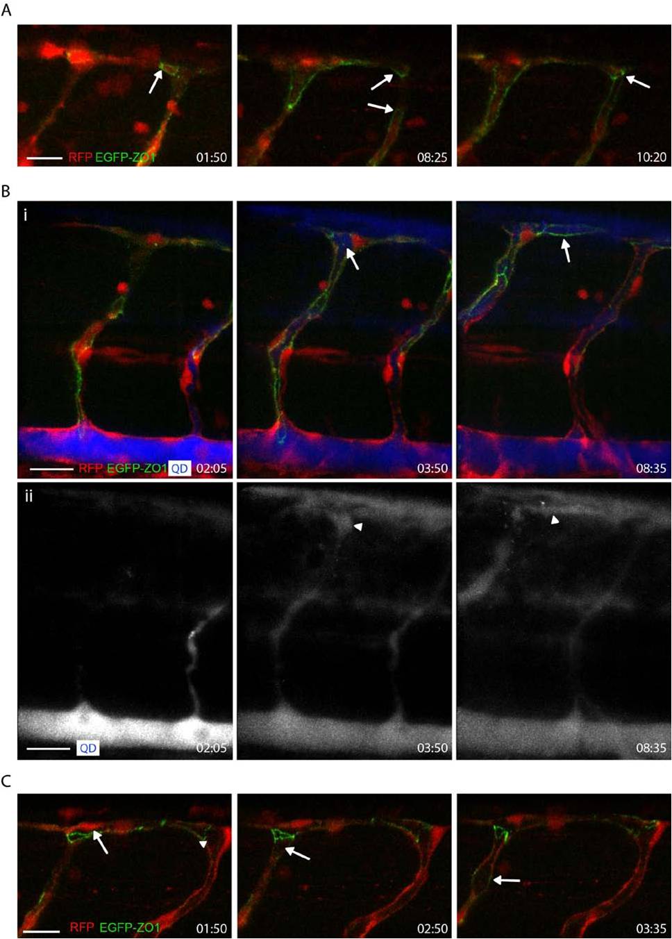
Lumen Invagination Visualized by QD Injection, Related to Figure 4 (A) Cell rearrangements do not depend on blood circulation. Series of pictures from movie S3. Fusion spots are transformed into rings (arrow at 01:50) and cell rearrangements (arrows at 08:25 and 10:20) still occur in sih morphant embryos.
(B) Microangiography in Tg(fli1ep:Gal4ffubs3; UAS:RFP; UAS:ZO1-EGFPubs5) embryos. Still pictures from movie S5. (i) show merge and (ii) show QD channel only. The SA to the left undergoes membrane invagination, similar to the events shown in Fig. 4. By invagination the lumen reaches the proximal site of the dorsal fusion point (arrow and arrowhead at 3:50). Anastomosis is completed when the luminal space within the fusion point becomes filled by QD (arrow and arrowhead at 8:35). (C) Dorsal to ventral lumen invagination. Still pictures from movie S6. The lumen extends from the DLAV (arrowhead) into the ISV (follow arrow from 01:50-03:35). The absence of longitudinal junctions indicates a transcellular lumen formation. The scale bar in all panels represents 20 μm.