
Fig. 3
- ID
- ZDB-IMAGE-111220-11
- Genes
- Publication
- Kimmel et al., 2011 - Requirement for Pdx1 in specification of latent endocrine progenitors in zebrafish
- All Figures
- Figures for Kimmel et al., 2011
|
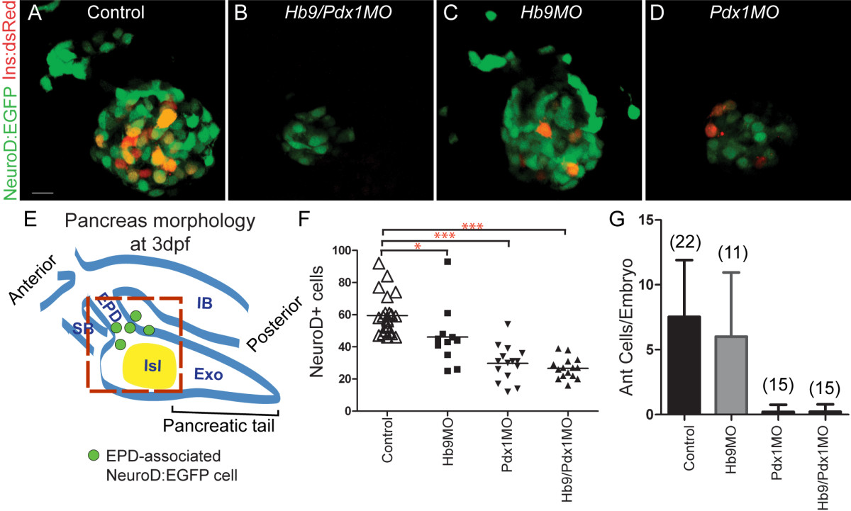
Fig. 3 Absence of ventral bud endocrine precursors in hb9/pdx1 and pdx1 morphant embryos. (A-D) Projections of confocal stacks showing native fluorescence of 3 days post fertilization (dpf) TgBAC(NeuroD:EGFP)nl1; Tg(ins:dsRed)m1018 embryos. In uninjected control embryos (A) and in hb9 morphants (C), enhanced green fluorescent protein (EGFP)+ cells are found in the islet and in smaller numbers also anterior to the islet. In hb9/pdx1 double morphants (B) and pdx1 morphants (D), the number of islet-associated EGFP+ cells is strongly reduced and anterior cells are missing. Few Ins:DsRed cells are present in pdx1 single morphants at 3 dpf (D), reflecting slow maturation of the DsRed fluorophore. (E) Schematic of pancreas morphology at 3 dpf. The principal islet (Isl) at this stage is primarily dorsal bud derived. The ventral bud contributes new NeuroD:EGFP cells (green circles), and generates exocrine pancreas (Exo) and extrapancreatic duct (EPD). IB, intestinal bulb; SB, swim bladder. Red box delineates region included for quantitation of islet and newly emerging anterior endocrine cells. (F) Quantitation of pancreatic EGFP+ cells (*P < 0.05, ***P < 0.001 with P values determined using one-way analysis of variance (ANOVA) with Bonferroni′s post test). (G) Quantitation of anteriorly positioned EGFP+ cells in uninjected and morpholino injected embryos (error bars indicate standard deviation from the mean). Scale bar = 10 μM.