
Image
|
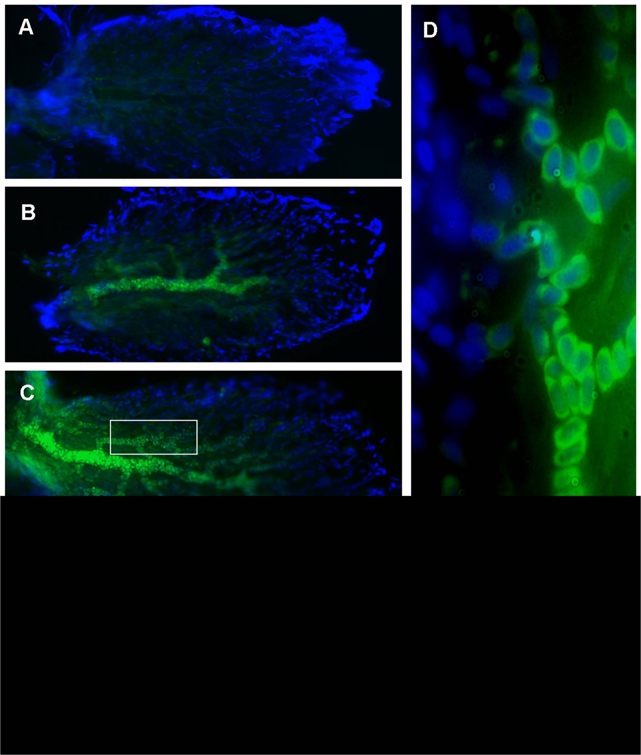
Figure Caption
Fig. 5 Expression of GH mRNA in zebrafish saccules.
Photomicrographs of saccules of zebrafish labeled with probes against GH mRNA via fluorescent in situ hybridization (FISH); green = FITC conjugated probe, blue = DAPI. Saccules from (A) a baseline control (not exposed to sound) and (B–E) sound-exposed zebrafish. (D) 100X image of the boxed area in (C), showing that GH mRNA appears to be localized perinuclearly in blood cells. (A–C) show only the caudal portion, while (E) shows a whole saccule. Caudal is to the right as in previous figures. Scale bars (D = 10 μm; E = 100 μm).
Acknowledgments
This image is the copyrighted work of the attributed author or publisher, and
ZFIN has permission only to display this image to its users.
Additional permissions should be obtained from the applicable author or publisher of the image.
Full text @ PLoS One