
Image
|
Figure Caption
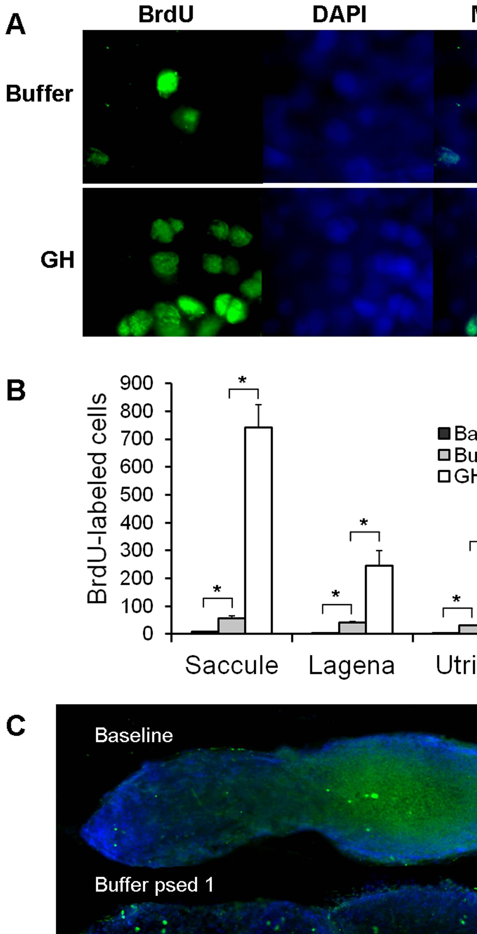
Fig. 3 Effect of GH on cell proliferation.
(A) 100X images of BrdU and DAPI labeling in the saccules of buffer- and GH-injected zebrafish. (B) Mean (±S.E.) number of BrdU-labeled cells in the saccules, lagenae, and utricles of baseline and buffer- or GH-injected zebrafish. N = 6; * P<0.001. (C) BrdU-labeling in the saccules of baseline, buffer- or GH-injected zebrafish at post-sound exposure day 1 (psed1). Scale bar = 100 μm. Rostral-caudal orientation is the same as Fig. 1A.
Acknowledgments
This image is the copyrighted work of the attributed author or publisher, and
ZFIN has permission only to display this image to its users.
Additional permissions should be obtained from the applicable author or publisher of the image.
Full text @ PLoS One