
Fig. 2
- ID
- ZDB-IMAGE-111215-26
- Publication
- Melville et al., 2011 - The feelgood mutation in zebrafish dysregulates COPII-dependent secretion of select extracellular matrix proteins in skeletal morphogenesis
- All Figures
- Figures for Melville et al., 2011
|
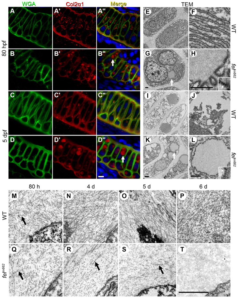
Fig. 2 Protein trafficking is disrupted in feelgood mutants. (A–D′′) Immunostaining of WGA (A–D) and Col2α1 (A2–D′) in the Meckel’s cartilage of 80-hpf (A–B′′) and 5-dpf (C–D′′) wild-type (WT; A-A′′, C-C′′) and feelgood. (B-B3, D-D3) embryos. Arrows in merged images indicate aberrant intracellular collagen localization in feelgood. Scale bar: 5 μm. (E–L) TEM images of 80-hpf (E–H) and 5-dpf (I–L) WT and feelgood chondrocytes. Arrows indicate distended ER membranes in feelgood cells. (M–T) TEM images of collagen fibrils in the extracellular space of feelgood mutants and WT siblings at 80 hpf and 4, 5 and 6 dpf. Arrows point to representative individual collagen fibrils. Scale bar: 1 μm.