
Image
|
Figure Caption
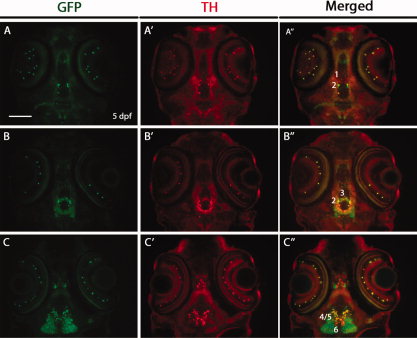
Fig. 7
Double immunostaining for green fluorescent protein (GFP) and tyrosine hydroxylase (TH) on horizontal cryosections of Tg(dat:EGFP) larvae at 5 days post-fertilization (dpf). The 5 dpf Tg(dat:EGFP) embryos were horizontally cryosectioned and stained with anti-GFP and anti-TH antibodies. GFP-positive cells and TH-positive cells are shown in green and red respectively. Numbers (1–6) indicate different groups of DA neurons in the ventral diencephalon. All sections are shown as anterior to the top. Scale bar = 100 μm.
Figure Data
Acknowledgments
This image is the copyrighted work of the attributed author or publisher, and
ZFIN has permission only to display this image to its users.
Additional permissions should be obtained from the applicable author or publisher of the image.
Full text @ Dev. Dyn.