
|
Fig. S3
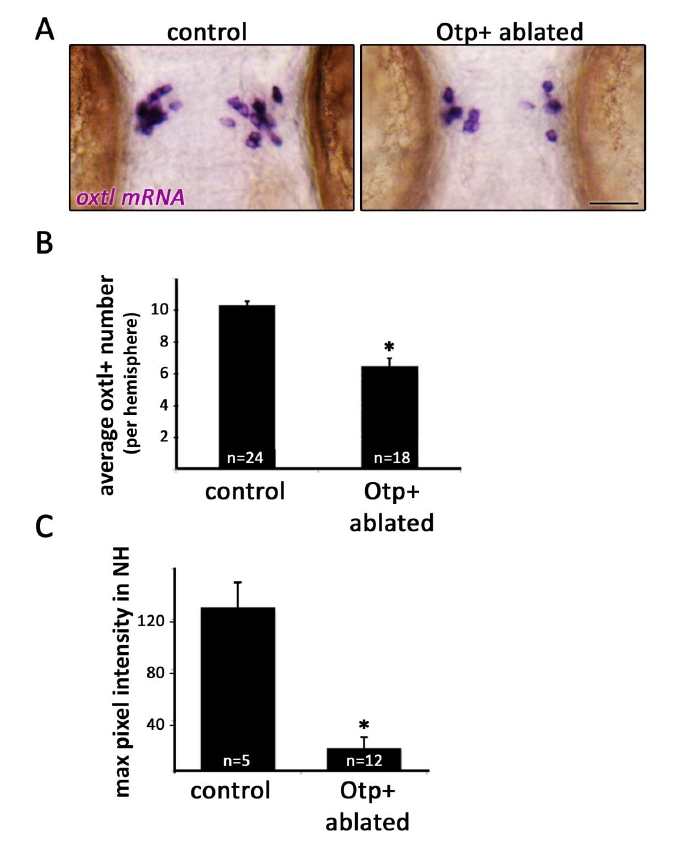
Figure S3 (related to Figure 5). Genetic cell ablation of Otp+ cells of the neurosecretory preoptic nucleus (NPO).
A,Micrographs of 3-day old embryos that underwent Otp+ cell ablation and their unablated siblings (see ‘RESULTS’ section). Embryos were subjected to in-situ hybridization with oxtl mRNA probe. Scale bar, 50μm.
B, Bar histogram depicting oxtl+ cell counts in control and Otpb+ cell ablated embryos. *p<0.001.
C, Oxtl protein was stained by immunofluorescence in 3-day old embryos that underwent Otp+ cell ablation and in their unablated siblings. Maximal pixel intensity (between 0-255) was recorded from confocal images of the NH region of each embryo and normalized for background pixel intensity. Oxtl staining was found to be reduced by ~85% in embryos that had undergone Otpb+ ablation. *p<0.001.
Reprinted from Developmental Cell, 21(4), Gutnick, A., Blechman, J., Kaslin, J., Herwig, L., Belting, H.G., Affolter, M., Bonkowsky, J.L., and Levkowitz, G., The hypothalamic neuropeptide oxytocin is required for formation of the neurovascular interface of the pituitary, 642-654, Copyright (2011) with permission from Elsevier. Full text @ Dev. Cell