
Image
|
Figure Caption
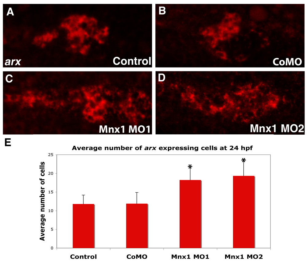
Fig. S6 Mnx1-deficient embryos have expanded expression of the arx alpha cell marker. (A-D) Whole-mount in situ hybridization for arx; fluorescent images of chromogenic in situ hybridizations. Ventral view of 24 hpf (A) uninjected control, (B) control MO-injected, (C) Mnx1 MO1-injected and (D) Mnx1 MO2-injected embryos. Anterior to left. (E) Mean (± s.d.) number of arx-expressing cells. *, P<0.001 (t-test, two-tailed distribution, Bonferroni correction), from a minimum of 11 embryos per group.
Figure Data
Acknowledgments
This image is the copyrighted work of the attributed author or publisher, and
ZFIN has permission only to display this image to its users.
Additional permissions should be obtained from the applicable author or publisher of the image.
Full text @ Development