
Fig. 1
- ID
- ZDB-IMAGE-111018-17
- Genes
- Publication
- Mouillesseaux et al., 2011 - Mutation in utp15 Disrupts Vascular Patterning in a p53-Dependent Manner in Zebrafish Embryos
- All Figures
- Figures for Mouillesseaux et al., 2011
|
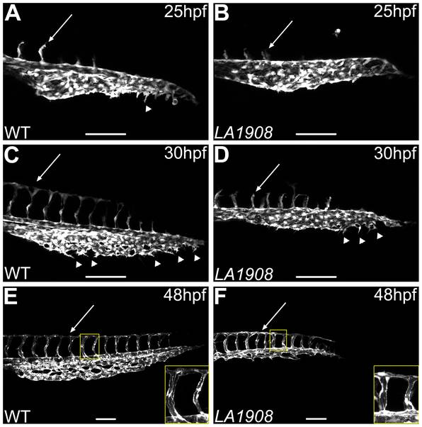
Fig. 1 Confocal microscopic analysis of angiogenesis in LA1908 embryos.
A–F, Confocal z-stack projection images were captured of the trunk and tail vasculature of wild type (A, C, E) and LA1908 mutant (B, D, F) embryos at 25 (A, B), 30 (C, D), or 48 hpf (E, F). CVP endothelial tip cells are indicated by arrowheads; extent of ISV maturation is indicated by arrows in A–D. Delay of primary ISV sprouting is most prominent at 30 hpf (C and D, arrows), when WT ISVs have reached their dorsal terminus and branched to form the DLAV (C), while mutant ISVs remain stunted (D). By 48 hpf, ISV number is equivalent between WT and mutant embryos and DLAV has formed in both groups (E, F, arrows). Defects in CVP angiogenesis result in a thinner and shorter plexus that lacks the complexity of WT embryos (F versus E). Insets in E and F, enlarged regions indicated by yellow rectangles, showing ISV lumens. Scale bars are 100 μm.