
Fig. 7
- ID
- ZDB-IMAGE-110909-73
- Publication
- Rothschild et al., 2011 - CaMK-II is a PKD2 target that promotes pronephric kidney development and stabilizes cilia
- All Figures
- Figures for Rothschild et al., 2011
|
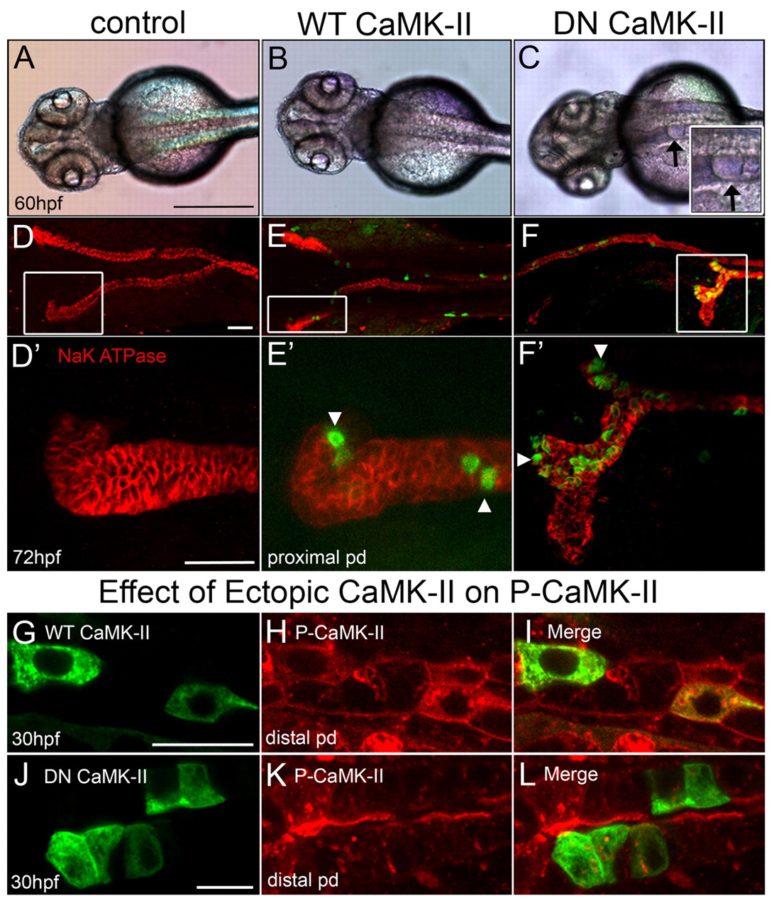
Fig. 7
Dominant-negative CaMK-II phenocopies camk2g1 morphants. (A-C) Dorsal views of 60 hpf embryos that were uninjected (control) or injected with kidney-targeted wild-type (WT) and dominant negative (DN) GFP-CaMK-II. Cysts are evident in DN CaMK-II embryos as shown in the inset (arrows). (D-F) Control and WT embryos immunostained for α1 Na+/K+-ATPase undergo proper convolution but DN embryos fail to convolute in proportion to expression (GFP). (D2-F2) Z-stack renderings of the anterior pronephric duct regions outlined by white boxes in D-F. (G-L) Kidney-targeted WT and DN CaMK-II are expressed in pronephric cells, but only DN CaMK-II diminishes P-CaMK-II. Scale bars: 500 μm in A; 20 μm in D,D2 10 μm in G,J.