
|
Fig. 5
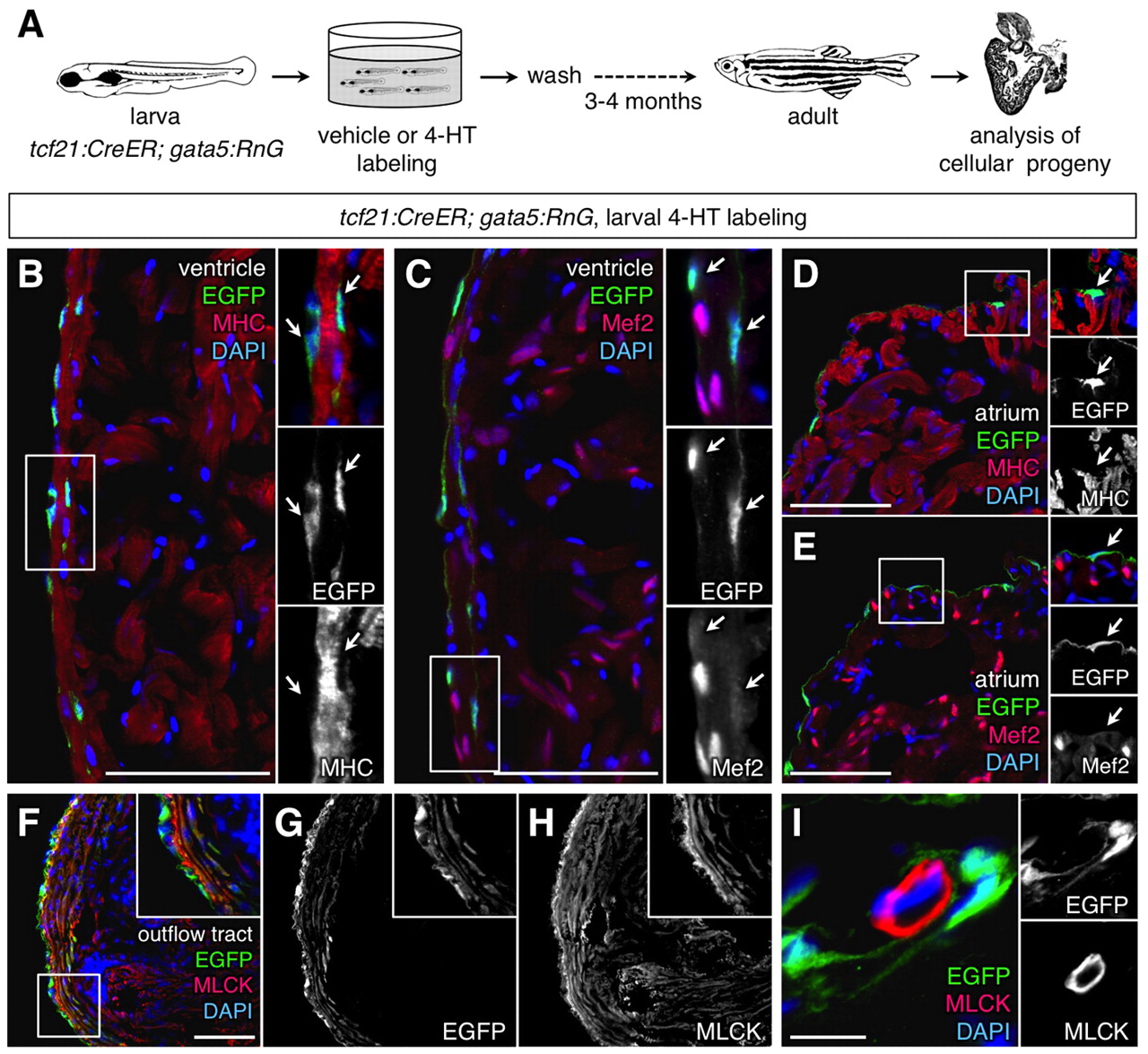
Adult progeny of labeled larval epicardial cells. (A) Schematic of the experimental design. (B,C) Portions of the ventricle from the lineage-traced heart, stained with an antibody for EGFP (green) and muscle markers (red). Lineage-labeled EGFP+ cells (arrows in inset) did not colocalize with cytosolic Myosin heavy chain (MHC; B) or nuclear Mef2 (C; n=10). The small EGFP+ region at the bottom of the inset of B is a thin strand of epicardial cytosol partially overlaying myocardial cytosol. A structure like this cannot be resolved by confocal imaging in processed tissue sections, but has neither the size nor morphology of a cardiomyocyte. Moreover, EGFP fluorescence was never observed in Mef2+ nuclei. (D,E) Portions of the atrium from the lineage-traced heart, stained with an antibody for EGFP (green) and muscle markers (red). Lineage-labeled EGFP+ cells (arrow in inset) did not colocalize with MHC (D) or Mef2 (E) (n=10). (F-I) EGFP+ cells in the outflow tract colocalize with Myosin light chain kinase (MLCK; red), a smooth muscle marker (F-H). This colocalization was not observed in coronary vascular smooth muscle (I). An antibody was used to detect EGFP in these experiments. Scale bars: 50 µm for B-H; 10 μm for I.