
|
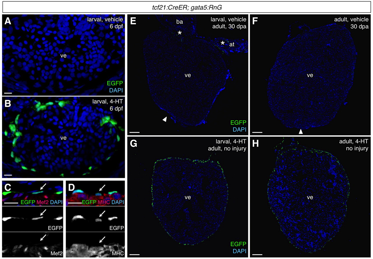
Fig. 4
Efficient and specific induced epicardial labeling in tcf21:CreER; gata5:RnG zebrafish. (A,B) tcf21:CreER; gata5:RnG larvae incubated with vehicle (A) or 4-HT (B) at 3-5 days post-fertilization (dpf) and visualized at 6 dpf, indicating 4-HT-induced labeling of the larval epicardium (green). Confocal projections of 10 μm z-stacks are shown. (C,D) Confirmation of epicardial specificity at 6 dpf embryos, assessed by Myosin heavy chain (MHC) and Mef2 staining. Arrows indicate an epicardial cell nucleus in each image. (E,F) 30 days post-amputation (dpa) ventricles from adult tcf21:CreER; gata5:RnG animals treated with vehicle as larvae (E) or adults (F). Occasional epicardial labeling (E, asterisks) was observed in injured or uninjured hearts. Arrowheads indicate apical regenerate. (G,H) 4-HT treatment at larvae or adult stages specifically labeled the majority of adult epicardial cells. at, atrium; ba, bulbus arteriosus; ve, ventricle. An antibody was used to detect EGFP in these experiments. Scale bars: 10 μm for A-D; 100 μm for E-H.