
|
Fig. 4
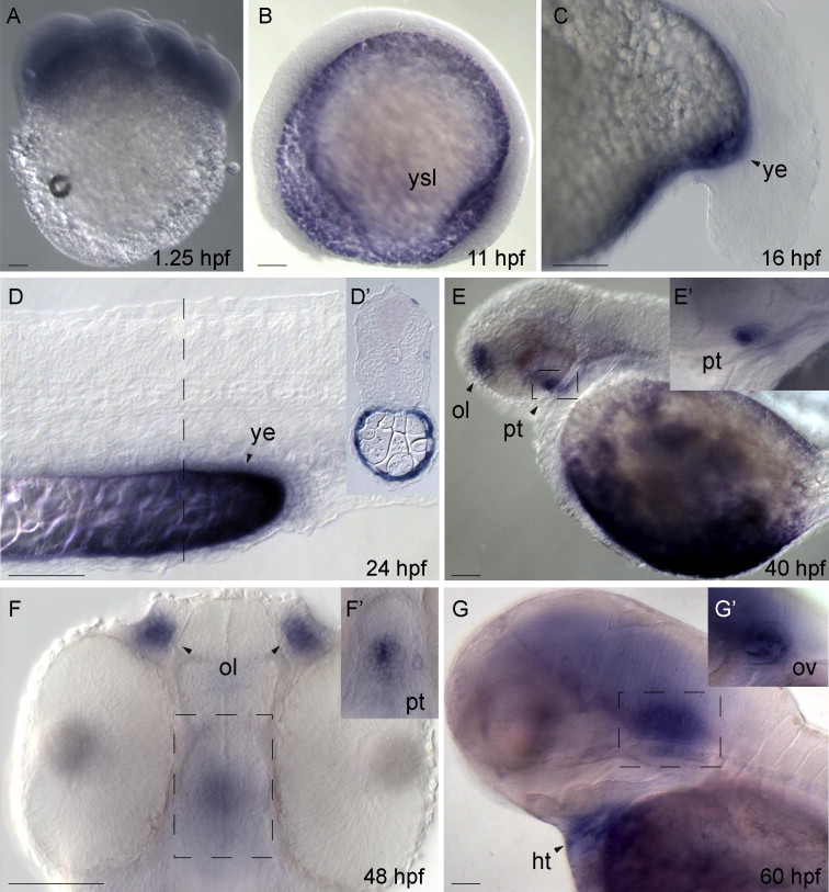
Developmental expression of soul1. (A), lateral view, animal pole to the top; (B–D, E, E2, G and G2) lateral view, anterior to the left; (D2) transverse resin section; (F and F2) ventral view, anterior to the top. Maternal soul1 mRNA is uniformly distributed at blastula stage (A). From 11 hpf, soul1 transcription is detected in the yolk syncytial layer (B), where its distribution becomes associated with the yolk extension as shown at 16 hpf (C) and 24 hpf (D and D2). Shown in (E and F) soul1 expression in the olfactory vesicles and pituitary gland (dotted box, enlarged in E2 and F2) at 40 and 48 hpf. In (G), soul1 expression in the heart and otic vesicles at 60 hpf (dotted box, enlarged in G2). ht, heart; pt, pituitary gland; ol, olfactory vesicle; ot, otic vesicle; ye, yolk extension; ysl, yolk syncytial layer. Black bars correspond to 100 μm.
Reprinted from Gene expression patterns : GEP, 11(5-6), Fortunato, A.E., Langellotto, F., and Sordino, P., Identification and expression of soul/p22HBP genes in zebrafish, 360-369, Copyright (2011) with permission from Elsevier. Full text @ Gene Expr. Patterns