
Fig. 4
- ID
- ZDB-IMAGE-110706-13
- Publication
- Chou et al., 2011 - Fascin 2b Is a Component of Stereocilia that Lengthens Actin-Based Protrusions
- All Figures
- Figures for Chou et al., 2011
|
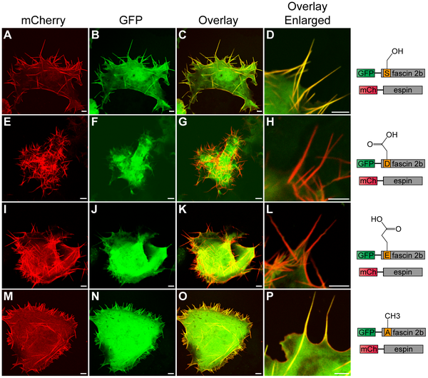
Fig. 4
Subcellular localization patterns of wild-type or phosphomutant fascin 2b proteins in live COS-7 cells that coexpress espin.
A cell (A–D) coexpressing GFP-WT fascin 2b and mCherry (mCh)-espin displays colocalization of these proteins in filopodia, which are detected as yellow in image overlays (C,D). Images of cells that coexpress mCherry-espin and either GFP-S38D fascin 2b (E–H) or GFP-S38E fascin 2b (I–L) reveal espin-laden protrusions (red) that lack the fascin 2b phosphomutant proteins (G,H,K,L). Micrographs of a cell that expresses both mCherry-espin and GFP-S38A fascin 2b (M–P) show that both proteins reside in filopodia (yellow) (O,P). Scale bars are 5 μm.