
Fig. 3
- ID
- ZDB-IMAGE-110622-117
- Publication
- Inoue et al., 2011 - One for all-a highly efficient and versatile method for fluorescent immunostaining in fish embryos
- All Figures
- Figures for Inoue et al., 2011
|
Fig. 3
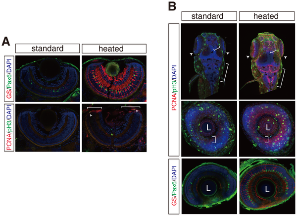
Multiple fluorescent immunostaining of cryosections and whole mount embryos by the heating method. (A) GS/Pax6 (top panels) and PCNA/phospho-histone H3 (pH 3) (bottom panels) immunostainings on cryosections of zebrafish retinae. GS and Pax6 immunostainings detect amacrine and Mueller glia cells. pH 3-positive mitotic cells (arrowheads) co-localize to PCNA-positive retinal progenitor cell region (brackets) in the ciliary marginal zone (CMZ). (B) Whole mount fluorescent immunostainings as in A with or without the heating method. Maximum projection of dorsal view of whole mount zebrafish (top panels). Coronal optical sections of zebrafish retina (middle and bottom panels). Brackets and arrowheads indicate proliferative zones of hindbrain, optic tectum, telencephalon, and CMZ. L; lens. Nuclei were counterstained with DAPI (blue).