
|
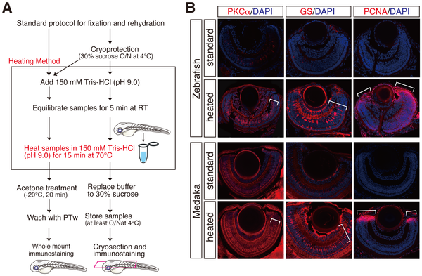
Fig. 1
Improvement of fluorescent immunostainings by a novel heating method. (A) Schematic flowchart including a novel heating step in the standard immunostaining for cryosections and whole mount preparations. Whole mount and cryoprotected fish embryos were heated at 70°C for 15 min in 150 mM Tris-HCl at pH 9.0 (framed), subjected to the respective standard protocol for immunostaining. Cryoprotected preparations also can be used for whole mount immunostaining (bifurcated arrows). (B) Fluorescent immunostainings with cryosections of zebrafish and medaka retinae using anti-PKCα, anti-GS and, anti-PCNA antibodies. PKCα, GS, and PCNA immunostainings mark bipolar, Mueller glia, and retinal progenitor cells, respectively. Note that the heating method (heated) but not standard method (standard) efficiently improved all the immunostainings (lower panels, see brackets). Nuclei were counterstained with DAPI (blue).