
Fig. 3
- ID
- ZDB-IMAGE-110527-3
- Genes
- Publication
- Krueger et al., 2011 - Flt1 acts as a negative regulator of tip cell formation and branching morphogenesis in the zebrafish embryo
- All Figures
- Figures for Krueger et al., 2011
|
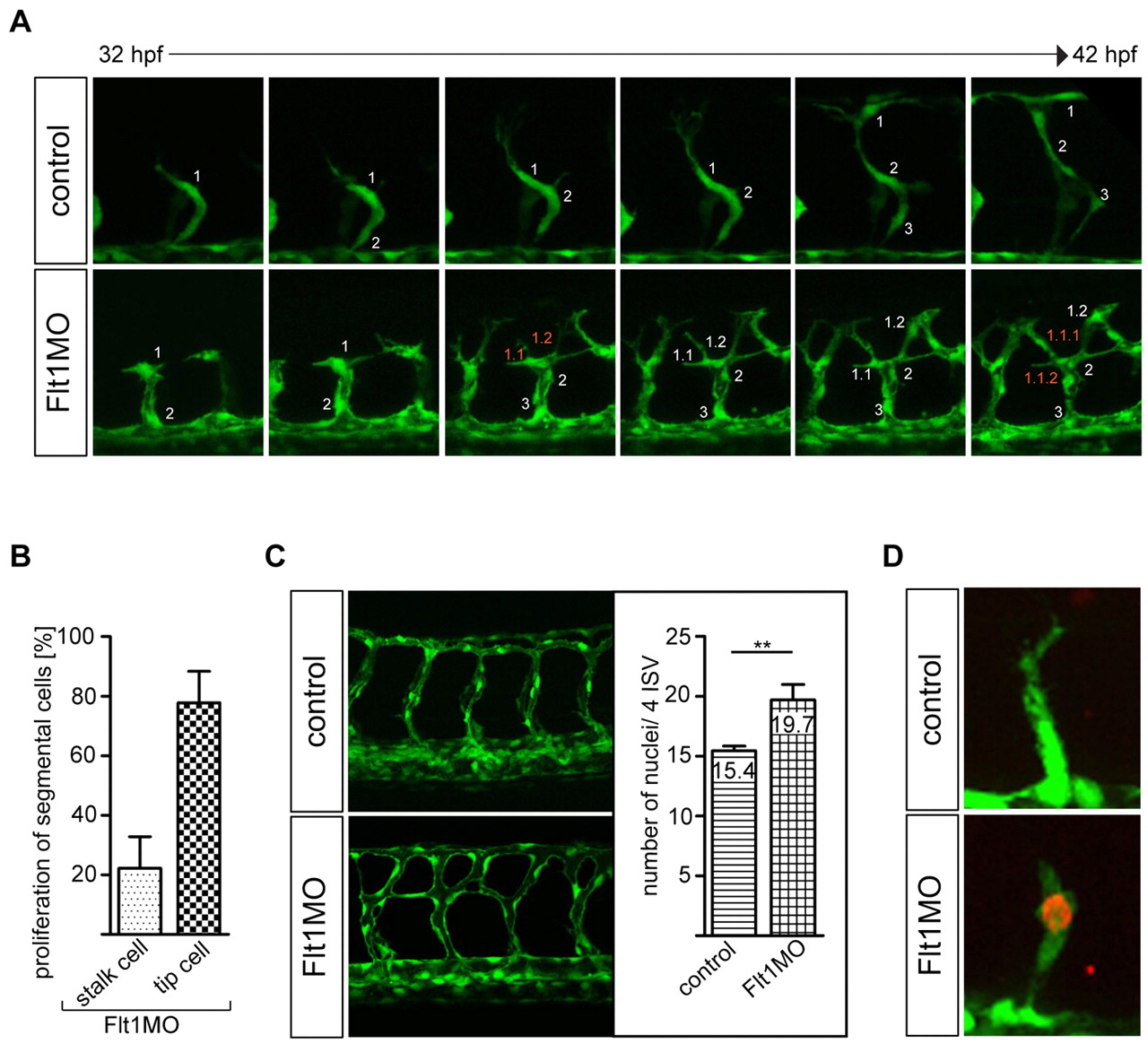
Fig. 3 Flt1 regulates endothelial tip cell formation. (A,B) Time-lapse in vivo imaging shows increased tip cell numbers and proliferating endothelial tip cells in segmental vessels of zebrafish flt1 morphants. Note that in flt1 morphants the leading cell number 1 gives rise to two progeny cells termed 1.1 and 1.2. y-axis shows the percentage of examined flt1 morphants with proliferation in tip or stalk cells. (C) Quantification of endothelial cell numbers in four consecutive segmental vessel (ISV) – dorsal longitudinal anastomotic vessel (DLAV) loops reveals an increase in flt1 morphants. (D) Anti-phospho-histone H3 staining confirms endothelial proliferation in segmental vessels of flt1 morphants. **, P<0.01; Student′s t-test. Error bars indicate s.e.m.