
Fig. s2
- ID
- ZDB-IMAGE-110429-9
- Publication
- González-Rosa et al., 2011 - Extensive scar formation and regression during heart regeneration after cryoinjury in zebrafish
- All Figures
- Figures for González-Rosa et al., 2011
|
Fig. s2
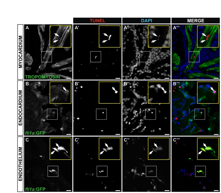
Cell death at 4 hours post-injury (4 hpi). (A-C222) Immunohistochemistry and TUNEL staining on sagittal sections of an adult Tg(fli1a:GFP) heart 4 hpi. Sections are the same as those shown in Fig. 4G-G222. Myocardial cells are revealed by staining for tropomyosin; endocardial and vascular endothelial cells by staining for GFP. Cell nuclei are stained with DAPI. Single channels and merged images are shown for representative TUNEL-positive cardiomyocyte (A-A222), endocardial cell (B-B222) and vascular endothelial cells (C-C222). Yellow insets show a higher magnification views of boxed areas. Arrows indicate TUNEL-positive nuclei. Scale bars: 10 μm.