
Fig. s1
- ID
- ZDB-IMAGE-110429-8
- Publication
- González-Rosa et al., 2011 - Extensive scar formation and regression during heart regeneration after cryoinjury in zebrafish
- All Figures
- Figures for González-Rosa et al., 2011
|
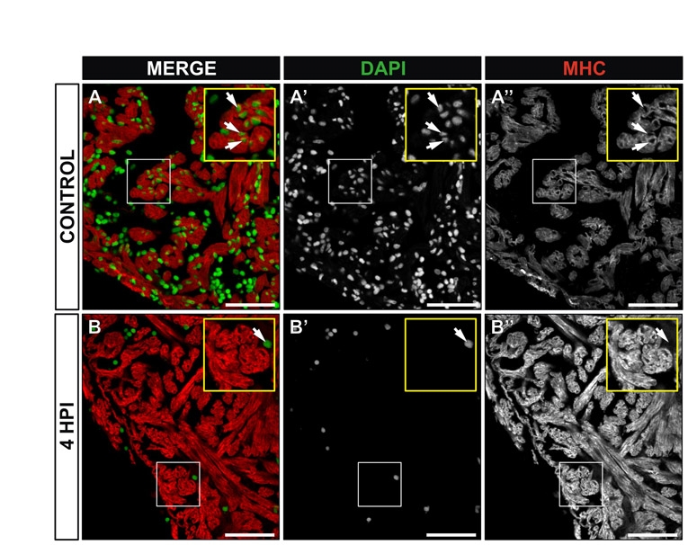
Fig. s1
Enucleated cardiomyocytes at 4 hpi. Immunohistochemistry on sections of control (A-A22) or cryoinjured zebrafish hearts at 4 hours post-injury (hpi) (B-B22). Myocardial cells are revealed by staining for myosin heavy chain (MHC, red) and cell nuclei are stained with DAPI (green). (A-A22) In control hearts, nucleated cardiomyocytes are detected in the compact and trabeculated myocardium (see magnified view in the inset). Arrows indiacte examples of nucleated cardiomyocytes. (B-B22) At 4 hpi, DAPI staining is lost to a great extent (B2), while MHC distribution is not affected (B22). Yellow insets show a higher magnification view of boxed area. A DAPI-positive cell in the lumen of the IA is shown (arrow). Note that neighboring cardiomyocytes do not contain nuclei. HPI, hours post-injury. Scale bars: 50 μm.