
Fig. 7
- ID
- ZDB-IMAGE-110429-7
- Publication
- González-Rosa et al., 2011 - Extensive scar formation and regression during heart regeneration after cryoinjury in zebrafish
- All Figures
- Figures for González-Rosa et al., 2011
|
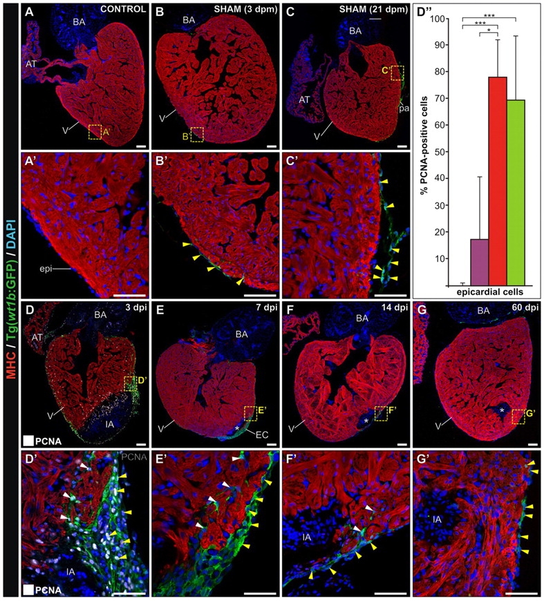
Fig. 7
Ventricular cryocauterization induces a proliferative response in epicardial cells. Immunostaining of sagittal Tg(wt1b:GFP) heart sections for myosin heavy chain (MHC) (red) and GFP (green). Nuclei are stained with DAPI (blue). Proliferating (PCNA-positive) cells are indicated with yellow arrowheads. Anterior is towards the top, ventral towards the right. Asterisks indicate the injured area (IA). Yellow arrowheads indicate GFP-positive cells in the epicardium. White arrowheads indicate GFP-positive cells in the compact layer. (A,A2) Untreated (control) heart revealing no GFP-positive cells. (B,B2) Sham-operated heart at 3 dpm; pericardial sac opening leads to GFP expression in epicardial cells. (C,C2) At 21 dpm, some GFP-positive cells are found in the epicardium. (D,D2) Cryoinjured heart at 3 dpi; proliferating (PCNA-positive) cells are shown white. Extensive proliferation is detected in the epicardium, especially at the borders of the IA. (D2) Nearly all GFP-positive cells are proliferating at this stage. The epicardial layer is several cell diameters thick. GFP-positive cells can also be found protruding into the compact layer. (D3) Quantification of proliferating epicardial cells in control sham-operated (violet) and cauterized hearts at regions close to (red) or distant from (green) the injured area at 3 dpm. For each condition, PCNA-positive cells were counted in three to five hearts (at least two sections per heart). Data are mean±s.d.; **P<0.01; ***P<0.001 (one-way ANOVA followed by Tukey′s honest significant difference test). (E,E2) At 7 dpi, GFP-positive epicardial cells cover the IA. (F,F2) Cryoinjured heart at 14 dpi; note the reduced thickness of the epicardial cap compared with earlier stages. (G,G2) Cryoinjured heart at 60 dpi. GFP-positive cells can also be found covering the remnants of the IA, although the epicardial layer has returned to its normal thickness of one cell diameter. AT, atrium; BA, bulbus arteriosus; dpi, days post-injury; dpm, days post-manipulation; epi, epicardium; IA, injured area; pa, pericardial adhesion. Scale bars: general view, 100 μm; higher magnification views, 50 μm.