
|
Fig. 6
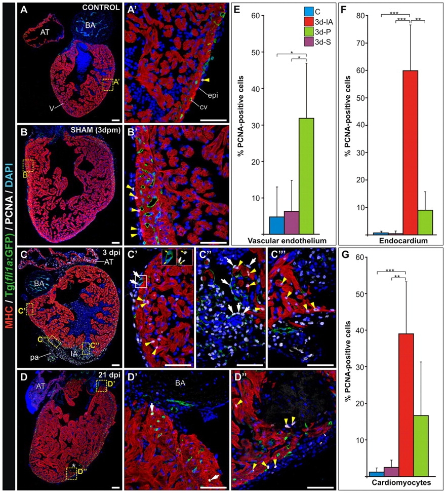
Ventricular cryocauterization induces a proliferative response in cardiac cells. Immunostaining of sagittal Tg(fli1a:GFP) heart sections for myosin heavy chain (MHC, red), GFP (green) and proliferating cell nuclear antigen (PCNA; white). Nuclei are stained with DAPI (blue). Proliferating (PCNA-positive) cells are indicated with yellow arrowheads. Anterior is towards the top, ventral towards the right. (A,A2) Untreated (control) heart revealing few proliferating cells. (B,B2) Sham-operated heart; pericardial sac opening leads to a limited increase in cell proliferation in the epicardium. (C-C4) Cryoinjured heart at 3 dpi. (C2) Extensive proliferation is detected away from the injured area (IA) in epicardium, cardiomyocytes and endocardial cells. White arrows indicate proliferating vascular endothelium. Inset shows a detailed view of proliferating endothelial cells (yellow arrows indicate PCNA-positive nuclei). (C3) In the trabeculated myocardium bordering the IA, both cardiomyocytes (yellow arrowheads) and endocardial cells (white arrows) are highly proliferating. (C23) PCNA-positive cardiomyocytes in the compact layer are indicated by yellow arrowheads. (D) Cryoinjured heart at 21 dpi. Asterisk indicates the IA. (D2) Only a few proliferating cells are detected in the periphery (white arrows indicate coronary endothelial cells). (D3) Few proliferating cells (yellow arrowheads) are found close to the IA. (E-G) Quantification of proliferating vascular endothelial cells, endocardial cells and cardiomyocytes in control (C), sham-operated (3d-S) and cauterized hearts at regions close to (3d-IA) or distant from (3d-P) the injured area at 3 dpm. For each condition, PCNA-positive cells were counted in three to five hearts (at least two sections per heart). Data are mean±s.d.; *P<0.05; **P<0.01; ***P<0.001 (one-way ANOVA followed by Tukey′s honest significant difference test). AT, atrium; BA, bulbus arteriosus; cv, coronary vessel; dpm, days post-manipulation; dpi, days post-injury; epi, epicardium; IA, injured area; pa, pericardial adhesion. Scale bars: general view, 100 μm; higher magnification views, 50 μm.