
Fig. 3
- ID
- ZDB-IMAGE-110429-3
- Publication
- González-Rosa et al., 2011 - Extensive scar formation and regression during heart regeneration after cryoinjury in zebrafish
- All Figures
- Figures for González-Rosa et al., 2011
|
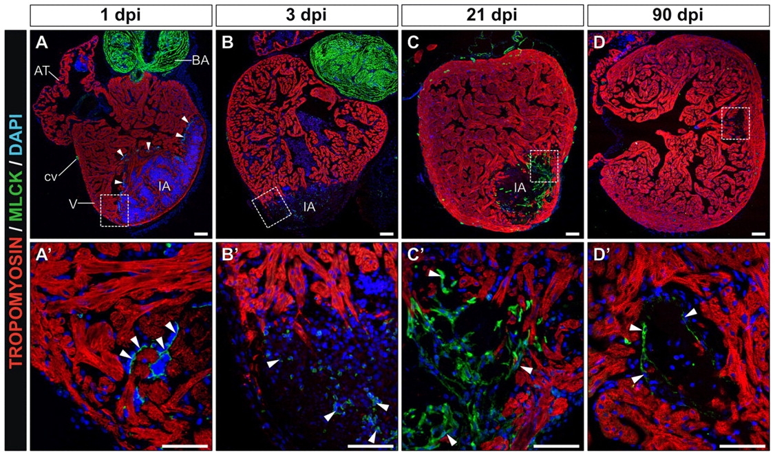
Fig. 3
Cryocauterization-induced myofibroblast accumulation and subsequent elimination during scar removal. (A-D) Immunohistochemistry on sagittal sections of cauterized hearts on the indicated days post-injury with antibodies to tropomyosin (red) and myosin light chain kinase (Mlck) (green); nuclei are stained with DAPI (blue). Anterior is towards the top, ventral towards the right. (A2-D2) Higher magnification views of the boxed areas in A-D. Arrowheads indicate Mlck-positive cells. (A,A2) Note the accumulation of Mlck at the borders of the injured region at 1 dpi, revealing activated thrombocytes. (B,B2) At 3 dpi, some Mlck-positive cells appear at the injury site. (C,C2) At 21 dpi, a Mlck-positive scar has formed, which partially persists until late stages of regeneration. (D,D2) At 90 dpi, only a few Mlck-positive cells can be detected in the scarred region. AT, atrium; BA, bulbus arteriosus; cv, coronary vessel; dpi, days post-injury; IA, injured area; V, ventricle. Scale bars: 100 μm.