
Fig. 2
- ID
- ZDB-IMAGE-110429-2
- Publication
- González-Rosa et al., 2011 - Extensive scar formation and regression during heart regeneration after cryoinjury in zebrafish
- All Figures
- Figures for González-Rosa et al., 2011
|
Fig. 2
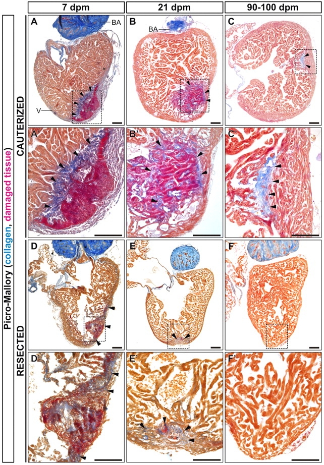
Comparison of collagen deposition and removal dynamics in cauterized and resected hearts. (A-F) Picro-Mallory stained sagittal sections of adult zebrafish heart fixed at the indicated days after cryocauterization (A-C) or resection (D-F) of the ventricular apex. Collagen is stained blue, damaged tissue in red and myocardium in brown. (A2-F2) Boxed areas of the damaged region shown at higher magnification. Anterior is towards the top, ventral towards the right. The strong collagen staining at the bulbus arteriosus acts as a positive control. (A-C2) Massive collagen deposition can be observed upon cauterization, which persists until late stages, indicating the formation of a scar. (D-F2) Resection triggers less collagen deposition than observed upon cryoinjury. Although a collagen scar persists until late stages after cauterization (C), it completely disappears after resection (F,F2). Arrowheads indicate sites of collagen deposition. BA, bulbus arteriosus; dpa, days post-amputation; dpi, days post-injury; IA, injured area; V, ventricle. Scale bars: 100 μm.