
IMAGE
Fig. S8
- ID
- ZDB-IMAGE-110428-20
- Publication
- Bussmann et al., 2011 - Arterial-venous network formation during brain vascularization involves hemodynamic regulation of chemokine signaling
- All Figures
- Figures for Bussmann et al., 2011
Image
|
Figure Caption
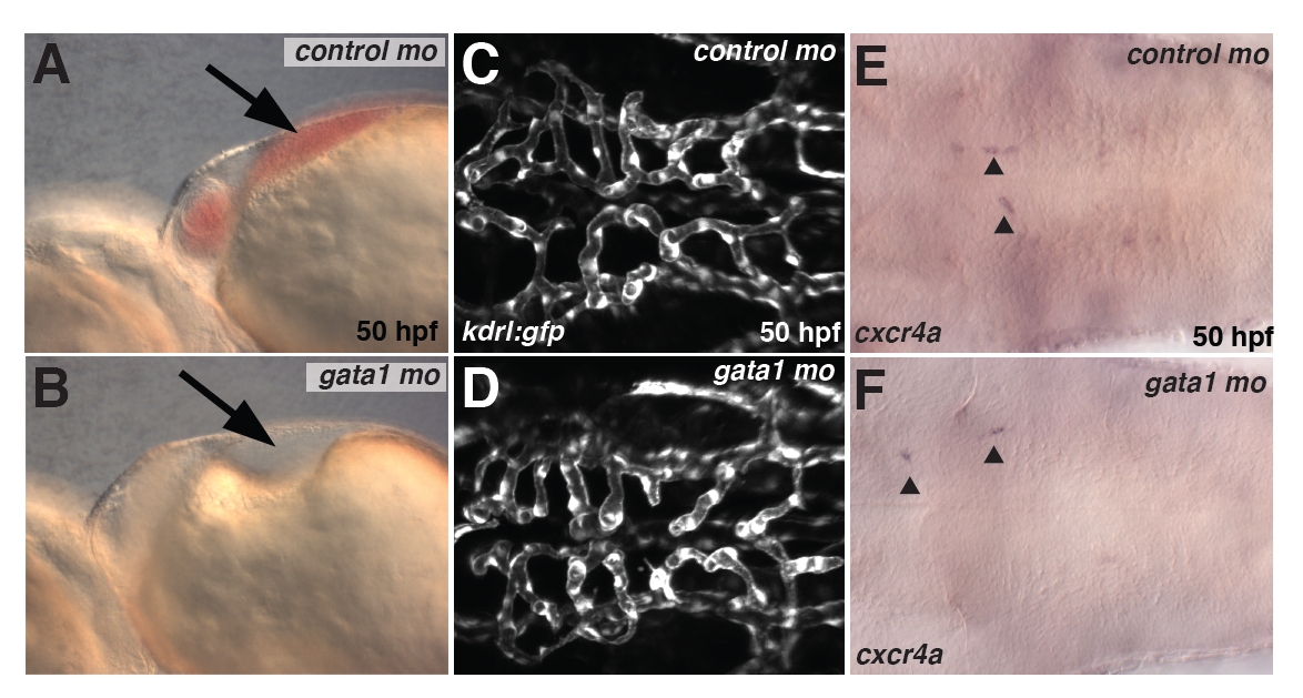
Fig. S8
Loss of erythrocytes does not affect hindbrain vascular patterning. (A,B) Brightfield image of embryos injected with control MO (A) or gata1 MO (B) at 50 hpf. Note the absence of erythrocytes in the heart region in gata1 MO-injected but not control MO-injected embryos (arrow). (C,D) Maximum projections of confocal z-stacks of kdrl:gfp transgenic embryos at 50 hpf injected with control MO (C) or gata1 MO (D) at 50 hpf. (E,F) Expression of cxcr4a in control MO-injected embryos (E) and gata1 MO-injected embryos (F).
Acknowledgments
This image is the copyrighted work of the attributed author or publisher, and
ZFIN has permission only to display this image to its users.
Additional permissions should be obtained from the applicable author or publisher of the image.
Full text @ Development