
Fig. 5
- ID
- ZDB-IMAGE-110407-15
- Publication
- Kim et al., 2011 - Zebrafish model of tuberous sclerosis complex reveals cell-autonomous and non-cell-autonomous functions of mutant tuberin
- All Figures
- Figures for Kim et al., 2011
|
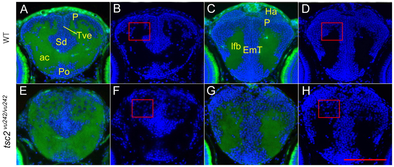
Fig. 5 Disruption of the gray and white matter in the anterior forebrain of tsc2vu242/vu242 mutants. Cross-sections through the anterior forebrain of wild-type larvae (A–D) and tsc2vu242/vu242 mutant larvae (E–H) at 7.5 dpf. A restricted telencephalon area containing gray and white matter was analyzed in wild-type (A,B) and tsc2vu242/vu242 mutant (E,F) larvae at 7.5 dpf. (B,F) DAPI-channel-alone image of A and E. The most anterior diencephalon of the wild-type is shown in C and mutant in G, with DAPI channel in D and H. Rectangles in F and H indicate disrupted pallium layers and ectopically mis-positioned cells within the white matter. Green color is tissue autofluorescence; blue is DAPI staining. P, pallium; Po, preoptic region; Sd, dorsal division of subpallium; Tve, telencephalic ventricle; ac, anterior commissure; Ha, habenula; EmT, eminentia thalami; lfb, lateral forebrain bundle. Scale bar: 100 μm.