
Fig. 9
- ID
- ZDB-IMAGE-110324-34
- Genes
- Antibodies
- Publication
- Sheets et al., 2011 - Ribeye is required for presynaptic CaV1.3a channel localization and afferent innervation of sensory hair cells
- All Figures
- Figures for Sheets et al., 2011
|
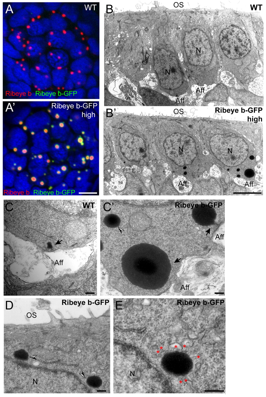
Fig. 9
Stable overexpression of Ribeye b in hair cells. (A,A2) Representative confocal z-projections at the basolateral end (~2.5 μm thick) of hairs cells in neuromast 2. Merged images include Ribeye b-GFP (green), Ribeye b (red) immunolabel and DAPI (blue). Scale bar: 3 μm. (B-E) Representative TEM sections through the ears of 4 dpf wild-type and transgenic larvae with high expression of Ribeye b-GFP. Scale bars: 2 μm in B; 250 nm in C-E. Aff, afferent terminal; N, nucleus; OS, otic space. (B,B2) Representative hair cells from wild-type and transgenic larvae. (C,C2) Representative ribbon synapses. Black arrows indicate attached ribbon synapses adjacent to afferent postsynaptic densities. Black and white arrowhead in C2 indicates an ectopic ribbon body. (D) Ectopic electron-dense aggregates (black and white arrowheads) at the apical end of the hair cell, next to the nucleus. (E) Magnified image of the lower aggregate in D. Red asterisks indicate attached vesicular structures.