
|
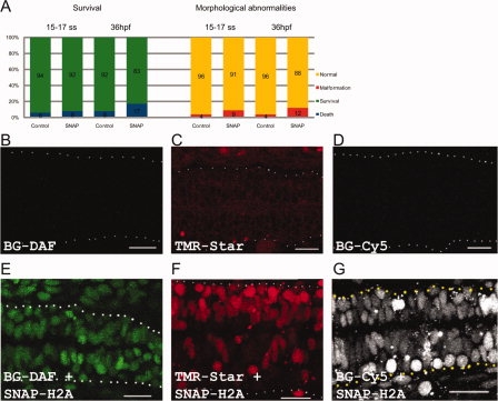
Fig. 1 H2A-SNAP labeling in live zebrafish embryos. A: Quantification and comparison of lethality and abnormal development between wild type and SNAP injected embryos at 15–17 somites and at 36 hr post-fertilization (hpf). B–D: Wild type embryos injected with (B) BG-DAF, (C) TMR Star, or (D) BG-Cy5 substrates. E–G: Confocal pictures of embryos injected with H2A-SNAP and (E) BG-DAF, (F) TMR Star, and (G) BG-Cy5. F: Embryo was labeled in a mosaic fashion, therefore nuclei on the right side of the midline are more strongly labeled than nuclei on the left side of this structure. B–D: Projection of 45 planes encompassing the spinal cord (dotted lines delimit the spinal cord). E–G: Projection of 5 planes in the spinal cord. B–G: 36-hpf embryos; anterior to the left. Scale bars = 20 μm.