
Fig. S3
- ID
- ZDB-IMAGE-110317-17
- Publication
- Osborn et al., 2011 - Cdkn1c drives muscle differentiation through a positive feedback loop with Myod
- All Figures
- Figures for Osborn et al., 2011
|
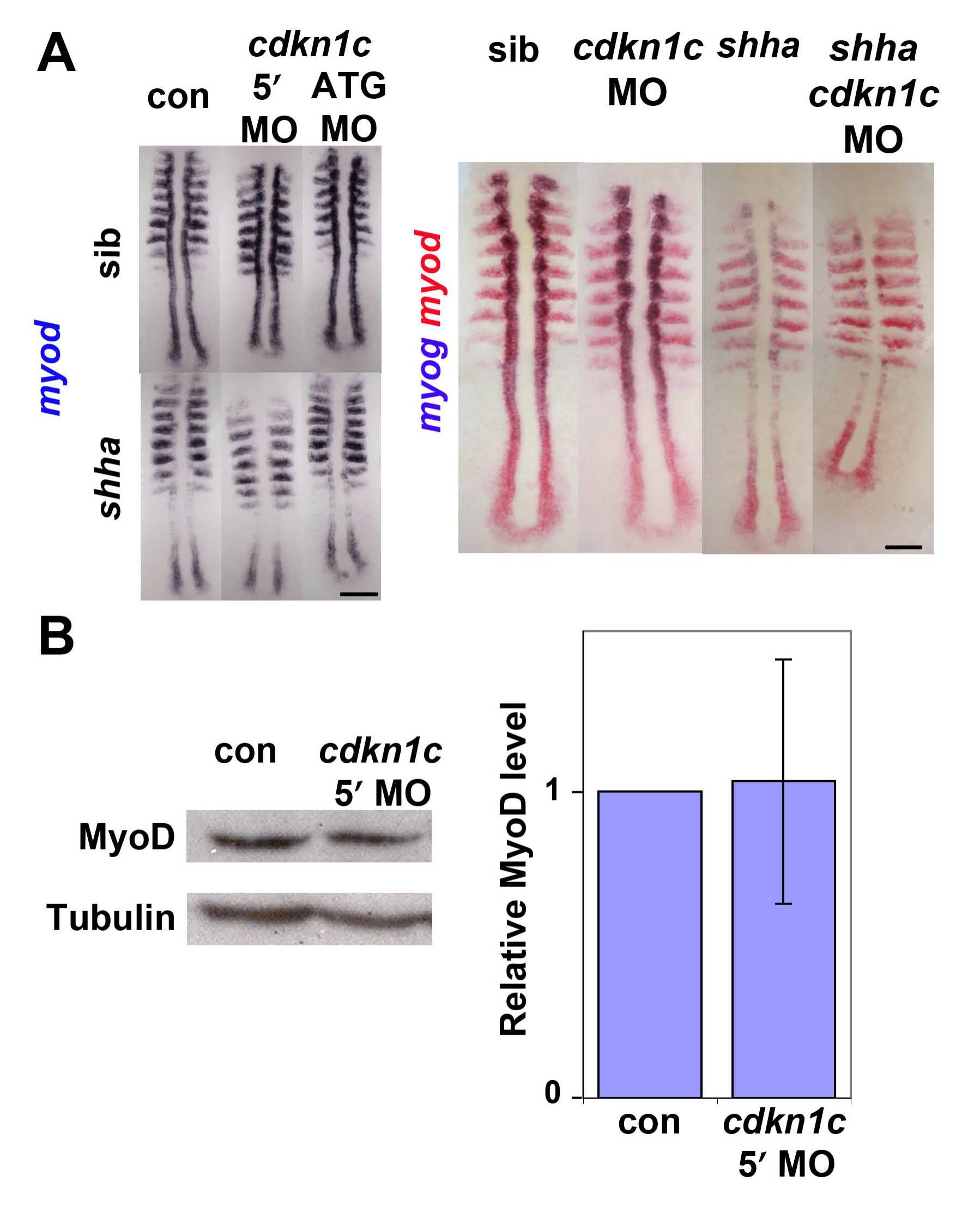
Fig. S3 Cdkn1c knockdown does not affect myod mRNA in presomitic mesoderm
(A). In situ mRNA hybridization for myod alone (left panel) or myod and myog (right panel) in a shhatbx392/+ incross injected with cdkn1c MOs or vehicle-control. Note the reduced numbers of myog-expressing cells in shha;cdkn1c MO embryo. Dorsal view of flatmounts at 8 som, anterior to top. (B). Western analysis of Myod in 24 hpf embryos injected with cdkn1c MO. A single major band of Mr ~ 44 000 is not reduced relative to β-Tubulin loading control (Mr ~ 55 000). When averaged over three experiments on 16–24 hpf embryos, no significant down-regulation of Myod band was detected after correction for protein loading. Bars: 25 μm.
Reprinted from Developmental Biology, 350(2), Osborn, D.P., Li, K., Hinits, Y., and Hughes, S.M., Cdkn1c drives muscle differentiation through a positive feedback loop with Myod, 464-475, Copyright (2011) with permission from Elsevier. Full text @ Dev. Biol.