
|
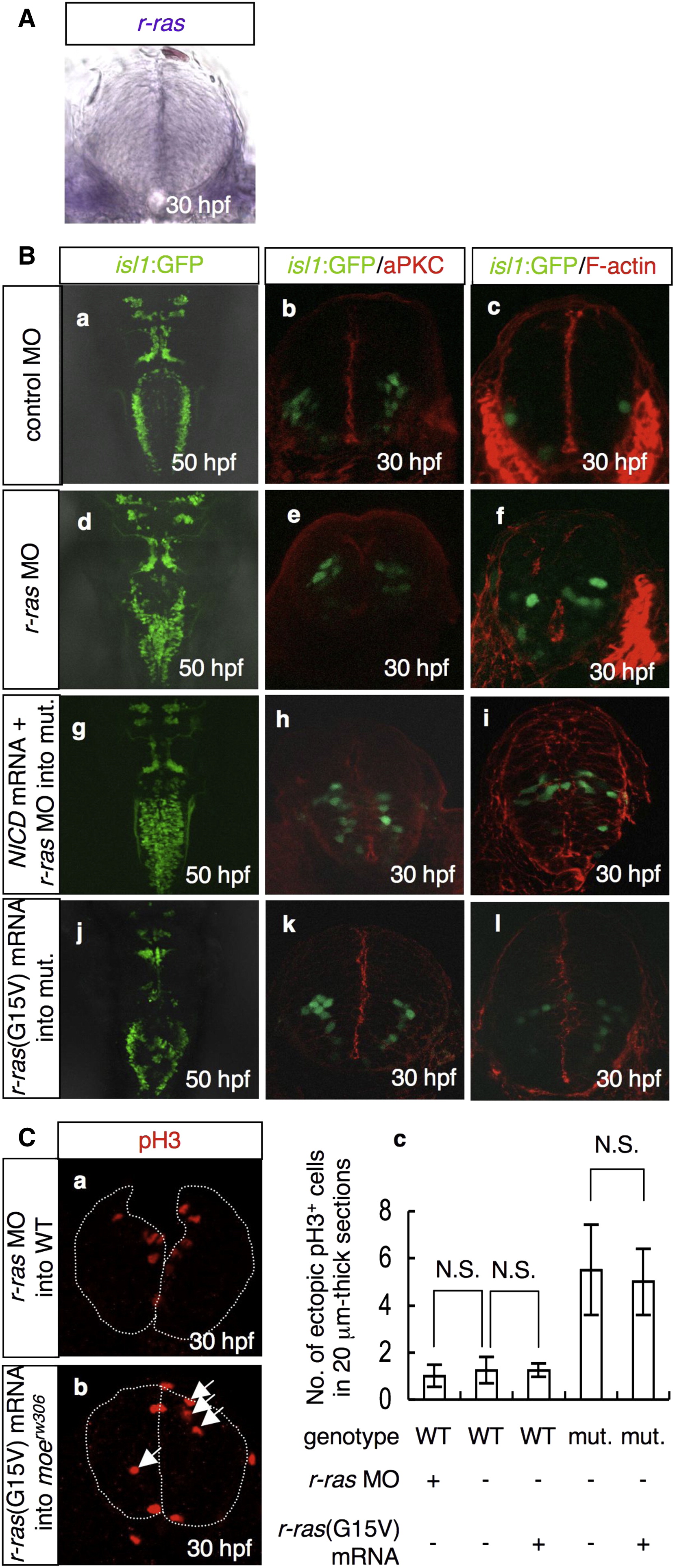
Fig. 7 R-Ras Functions Downstream of Moe and Notch to Maintain Neuroepithelial Polarity
(A) In situ hybridization for r-ras mRNA at 30 hpf in the WT caudal hindbrain. Cross-sectional view, dorsal to the top.
(B) The formation of the vagus motor nuclei (a, d, g, and j, dorsal view, 50 hpf), neuroepithelial polarity (b, e, h, and k; aPKC stain, red; cross-sectional views; 30 hpf), and intercellular junctions (c, f, i, and l; F-actin stain, red; cross-sectional views; 30 hpf) were examined in the isl1:GFP embryos injected with the control MO (a–c; 3.5 mg/ml) or r-ras MO (d–f; 3.5 mg/ml) and in the moerw306 mutants (mut.) injected with NICD FL mRNA (60 μg/ml) plus r-ras MO (g–i; 3.5 mg/ml) or r-ras(G15V) mRNA (j–l; 60 μg/ml). The vagus motor neurons are apparent in green (a–l).
(C) The WT (a and c) and moerw306 mutant (b and c) embryos were injected with r-ras MO (a and c) and r-ras(G15V) mRNA (b and c) and stained with anti-pH3 antibody. The number of ectopically dividing cells was assessed in 20 μm thick sections (c). The data shown are mean ± SEM; N.S. is an abbreviation for not significant.