
Fig. 2
- ID
- ZDB-IMAGE-110214-35
- Genes
- Antibodies
- Publication
- Ravanelli et al., 2011 - The Actin Nucleator Cordon-bleu is Required for Development of Motile Cilia in Zebrafish
- All Figures
- Figures for Ravanelli et al., 2011
|
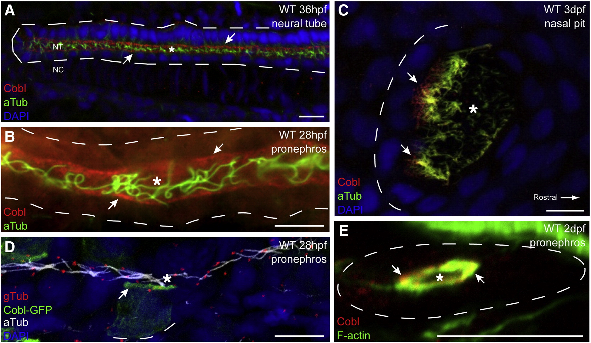
Fig. 2 Apical localization of Cobl protein in ciliated epithelial tissues.
(A) Whole mount confocal image of the caudal neural tube at 36 h post fertilization. Anti-Cobl staining reveals apical localization of Cobl in ciliated cells. Cobl is localized as distinct apical puncta, visible in the pronephric ducts at 28 hpf (B), and in the nasal placodes at 3 dpf (C). (D) mCoblGFP fusion proteins are able to concentrate apically in tissues where zfcobl is normally expressed. (E) Immunohistochemistry on transverse cryosections shows that Cobl localizes closely with apical F-actin in the pronephric duct. Arrows point to apical Cobl puncta. Asterisks denote lumens. Dashed lines demark basal laminae. F-actin (phalloidin), cilia (aTub, anti-acetylated tubulin), basal bodies (gTub, anti-gamma tubulin).
Reprinted from Developmental Biology, 350(1), Ravanelli, A.M., and Klingensmith, J., The Actin Nucleator Cordon-bleu is Required for Development of Motile Cilia in Zebrafish, 101-111, Copyright (2011) with permission from Elsevier. Full text @ Dev. Biol.