
Fig. 1
- ID
- ZDB-IMAGE-110111-22
- Genes
- Antibodies
- Publication
- Tsujimura et al., 2010 - A single enhancer regulating the differential expression of duplicated red-sensitive opsin genes in zebrafish
- All Figures
- Figures for Tsujimura et al., 2010
|
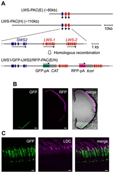
Fig. 1 Recapitulation of the LWS-1 and LWS-2 expression in the zebrafish retina by the fluorescent markers in the PAC clones.
(A) Construction of the LWS1/GFP-LWS2/RFP-PAC clones. The two PAC clones, LWS-PAC(E) and LWS-PAC(H), both encompass the two red opsin genes, LWS-1 and LWS-2, in addition to the blue opsin gene, SWS2. In the expanded view, the blue and red boxes indicate the exons of the blue and red opsin genes, respectively. The orientation of transcription is given by the arrows for each gene. In both the LWS1/GFP-LWS2/RFP-PAC(E) and LWS1/GFP-LWS2/RFP-PAC(H) clones, the first exons of LWS-1 and LWS-2 were replaced with the GFP-polyA-CAT and RFP-polyA-Kmr cassettes by site-specific homologous recombination, respectively. (B) A transverse section of the retina of an adult Tg(LWS1/GFP-LWS2/RFP-PAC(E))#1229 fish. The dorsal side is oriented at the top of each panel and the ventral side is at the bottom. GFP (green) is expressed in the ventral region (left), whereas RFP (magenta) is expressed in the central to dorsal region (middle). The right panel shows the merge of the left two panels with the transmitted light image taken by the differential interference contrast microscopy (DIC image). (C) A vertical section of the photoreceptor layer in the ventral retina of an adult Tg(LWS1/GFP-LWS2/RFP-PAC(H))#430 fish. The outer segments of LDCs are immunostained with an antibody against the zebrafish red opsin (magenta). Note that the GFP (green) is found mostly in the cell body of LDCs. Scale bars = 100 μm for (B), 10 μm for (C).