
Fig. 7
- ID
- ZDB-IMAGE-110105-7
- Genes
- Publication
- Seebald et al., 2011 - Zebrafish eve1 regulates the lateral and ventral fates of mesodermal progenitor cells at the onset of gastrulation
- All Figures
- Figures for Seebald et al., 2011
|
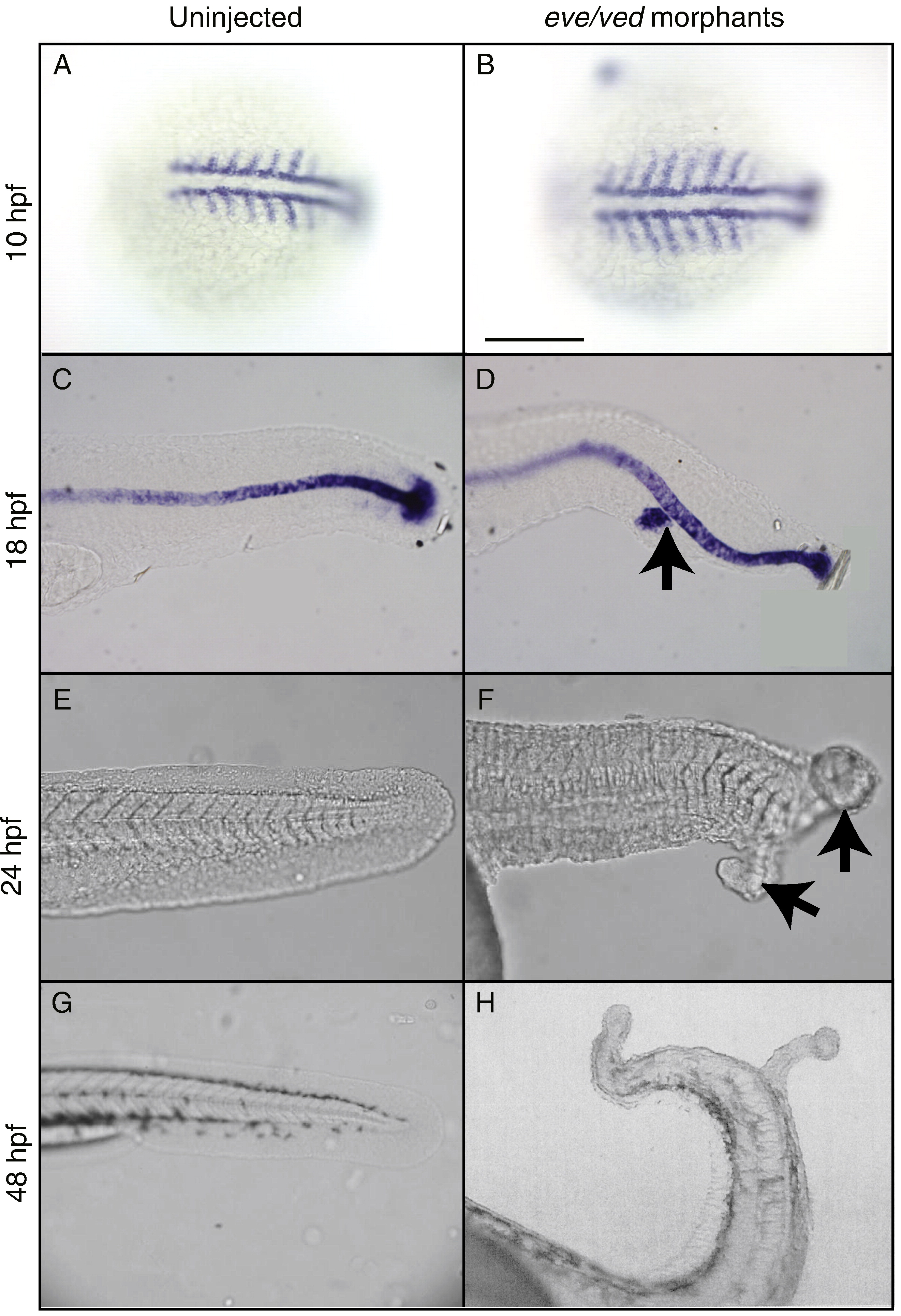
Fig. 7 Characterization of ectopic tail phenotypes of eve/ved morphant embryos by whole-mount in situ hybridization and morphological observation. A and B) Dorsal views with anterior to the left, showing myoD expression is normal in uninjected (A) (n = 15/15) and laterally expanded in eve/ved morphant (B) embryos at 10hpf (n = 23/25). C and D) Lateral views with anterior to the left, showing the ntl expression is normal in uninjected (C) (n = 18/18) and at an ectopic location (black arrow) in eve/ved morphant (D) embryos at 18 hpf (n = 10/50). E–H) Lateral views with anterior to the left, showing the tail (posterior body) of uninjected (E) and primary and secondary tails (black arrows) of eve/ved morphant (F) at 24 hpf (n = 9/50). G and H) Normal tail (posterior body) formation of uninjected (G) and ectopic tail formation of eve/ved morphant (H) embryos at 48 hpf. Scale bar, 200 μm.
Reprinted from Developmental Biology, 349(1), Seebald, J.L., and Szeto, D.P., Zebrafish eve1 regulates the lateral and ventral fates of mesodermal progenitor cells at the onset of gastrulation, 78-89, Copyright (2011) with permission from Elsevier. Full text @ Dev. Biol.