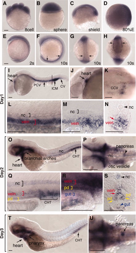
Fig. 4 Expression of par1 during zebrafish embryogenesis. A–V: Whole-mount in situ hybridization was performed on embryos at various stages: eight-cell (A); sphere (B); gastrula (C–E); 10-somite (F–H, arrows indicate posterior and ventral mesoderm); 1 days postfertilization (dpf; I–N; I: lateral view of a embryo showing par1 expression in the heart, posterior cardinal vein [PCV], cardinal vein [CV], intermediate cell mass of mesoderm [ICM]; J: high-magnification image of anterior region of I; K: dorsal view of the anterior region of the embryo showing the par1 expression in common cardinal vein [CCV]; L: high-magnification image of the trunk region of I, location of the notochord (nc) is indicated; M,N: sagittal and transverse sections of the trunk area indicated by the black line shown in I); day 2 (O–S; O: Lateral view showing par1 expression in the heart, branchial arches, caudal hematopoietic tissue [CHT]; P: dorsal view of the anterior region of O at high magnification; Q,R: lateral view of the trunk region of O at high magnification; S: transverse section at the trunk region showing par1 expression in notochord [nc], vein, pronephric duct [pd], and gut); 3 dpf (T–U; T: lateral view showing par1 expression in the heart, pharynx, CHT; U: dorsal view of the anterior region at high magnification; par1 expression in pancreas is indicated). Most embryos are shown in lateral view, with the animal pole up. Exceptions include G (dorsal view), H (dorsal–posterior view), and K, P, U (dorsal view). Scale bars = 100 μm.
Image
Figure Caption
Figure Data
Acknowledgments
This image is the copyrighted work of the attributed author or publisher, and
ZFIN has permission only to display this image to its users.
Additional permissions should be obtained from the applicable author or publisher of the image.
Full text @ Dev. Dyn.

