
|
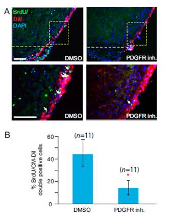
Fig. S6 PDGF signaling is essential for proliferation of epicardial cells during heart regeneration. Treatment of regenerating hearts with DMSO as a control or with PDGFR inhibitor (inh.). The hearts were treated from 2–7 dpa and collected at 7 dpa. (A) Epicardium of zebrafish hearts was labeled with Cell Tracker CMDiI at 2 dpa (BrdU, green; CM-DiI, red). The dashed line marks the approximate position of the amputation plane. BrdU-positive epicardial cells near the wound (marked by arrows) were quantified. (Scale bars = 50 μm.) (B) Statistical analysis of in vivo BrdU incorporation in epicardial cells during heart regeneration. BrdU-positive cells in the boxed area in A were quantified. Eleven hearts (n = 11) were analyzed in each group. Error bars indicate SEM. *P < 0.01.