
IMAGE
Fig. S2
- ID
- ZDB-IMAGE-101118-26
- Genes
- Publication
- Schuster et al., 2010 - Glial cell line-derived neurotrophic factor defines the path of developing and regenerating axons in the lateral line system of zebrafish
- All Figures
- Figures for Schuster et al., 2010
Image
|
Figure Caption
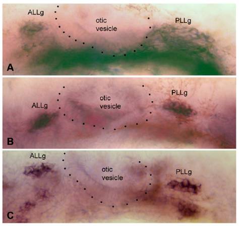
Fig. S2 Expression of gfr1a, gfr1b, and ret1 in the anterior (ALLg) and posterior (PLLg) lateral line ganglia, on either side of the otic vesicle (dotted outline), in 35-hpf embryos. C has been assembled from two slightly different focal planes.
Figure Data
Acknowledgments
This image is the copyrighted work of the attributed author or publisher, and
ZFIN has permission only to display this image to its users.
Additional permissions should be obtained from the applicable author or publisher of the image.
Full text @ Proc. Natl. Acad. Sci. USA